


30 June 2026
Cryo-EM helps identify the mechanisms of dental plaque formation

Periodontal (gum) disease is one of the most prevalent diseases worldwide, caused by the bacterium Porphyromonas gingivalis (P. gingivalis). In Japan alone, approximately 80% of adults over 30 years old are affected or considered at risk.
Published in Communications Biology, a joint study by the Okinawa Institute of Science and Technology (OIST), Tottori University, Hiroshima University, and Nagasaki University provides new insights into how this bacterium causes plaque formation. Using cryo-electron (cryo-EM) microscopy, they reveal the 3D structure of Mfa pili, an arm-like filament which enables the bacteria to stick to host tissues and other microbes.
First author Dr Satoshi Shibata, a former researcher in OIST’s Molecular Cryo-Electron Microscopy Unit and now Lecturer at Tottori University says, “By understanding how P. gingivalis attaches to host tissues, establishes infection, and participates in biofilm formation, we can inform the development of future therapeutic strategies. Our detailed structural information may serve as a drug-design template for identifying compounds that block attachment and infection.”
How P. gingivalis forms plaques
To attach to hosts or other microbes, P. gingivalis uses two different types of filaments (pili): Fim and Mfa. Both are made of multiple protein subunits, which join together to form long arm-like structures, and can bind to different types of bacteria and human tissues. In Mfa, most of these subunits are Mfa1 proteins.
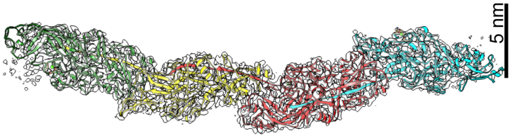
The authors have long been interested in the structure and function of these filaments, previously describing the structure of FimA, a key component of Fim pili. With this new publication they provide a fuller picture, elucidating the structure of Mfa1 chains central to Mfa pili and describing how these assemble and bind to other bacteria.
To understand filament formation, the researchers first polymerized the Mfa1 protein in vitro and analyzed it by cryo-EM, determining the structure at a near-atomic resolution of 3.0 Å.
By modifying the proteins, the team explored the role and importance of particular sites to filament assembly. They demonstrated how Mfa proteins come together in a process called strand exchange, dependent on interactions within one particular region of the protein (the C-terminus) for structural stability.
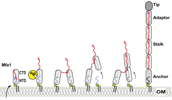
The cryo-EM mapping revealed metal ions within the Mfa filament, which, through further analysis, were identified as calcium. “Our tests suggest that this calcium binding can help the bacterium to avoid immune recognition,” highlights Shibata.
Using computer simulations, the researchers were also able to visualize the interaction of the Mfa filaments with Streptococcus gordonii, another bacterium which commonly binds to P. gingivalis in dental plaques. By identifying how these bacteria interact, scientists may more easily identify compounds to block these interactions, inhibiting plaque formation.
In addition to gum disease, P. gingivalis has been implicated in a wide variety of conditions, including pneumonia, diabetes, Alzheimer's disease, rheumatoid arthritis, stroke, cardiovascular disease, and adverse pregnancy outcomes. By providing detailed structural information, the researchers hope to aid scientists in developing treatments for P. gingivalis-related diseases.
Article Information
Specialties
Research Unit
Submit a press inquiry


