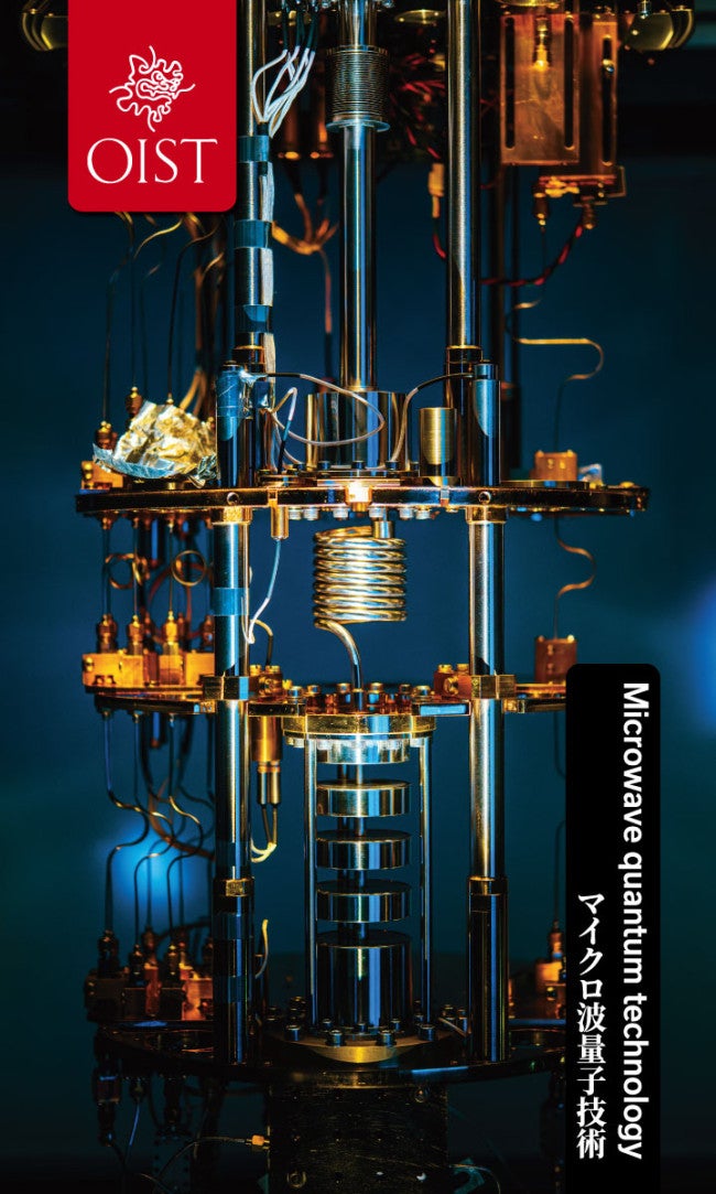
Researchers at OIST are developing an ultra-low-noise amplifier that uses the defects in the crystal structure of gemstones to boost extremely weak microwave signals. Beyond applications in ultra-sensitive magnetic resonance spectroscopy, this new amplifier could transform the field of cryogenic microwave quantum technologies and lead to the creation of scalable quantum computing hardware systems
Photo: Jeffery Prine
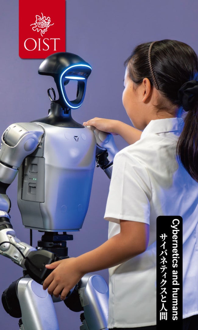
Cybernetics and Humans
Researchers from OIST and Sony CSL depict their work on robot-human coordination through synchronized dance moves. By studying how people and robots perceive, think, and move together, they aim to better understand the sense of 'self' and the evolving relationship between humans and computers that is shaping modern humanity
Photo: Jeffery Prine
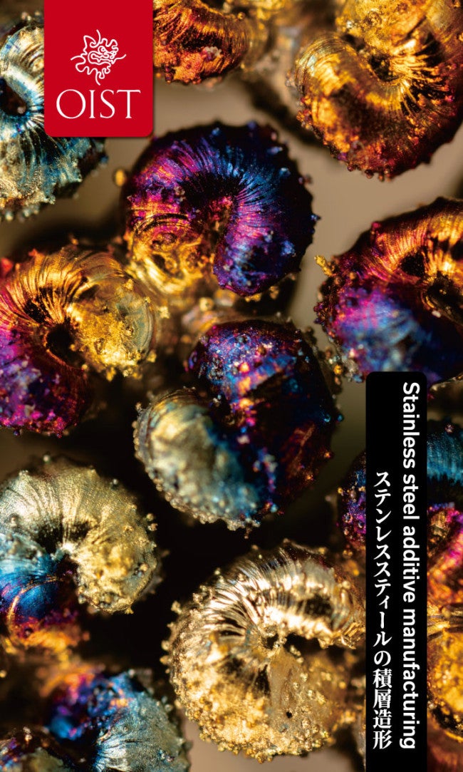
Stainless steel additive manufacturing
A magnified view of a 3D-printed stainless-steel support structure — a temporary framework that supports the object during printing. Oxidation patterns and unmelted metal particles create a unique and intricate landscape, showcasing the beauty and complexity achieveable through additive manufacturing (3D printing).
Photo: Michael Grunwald
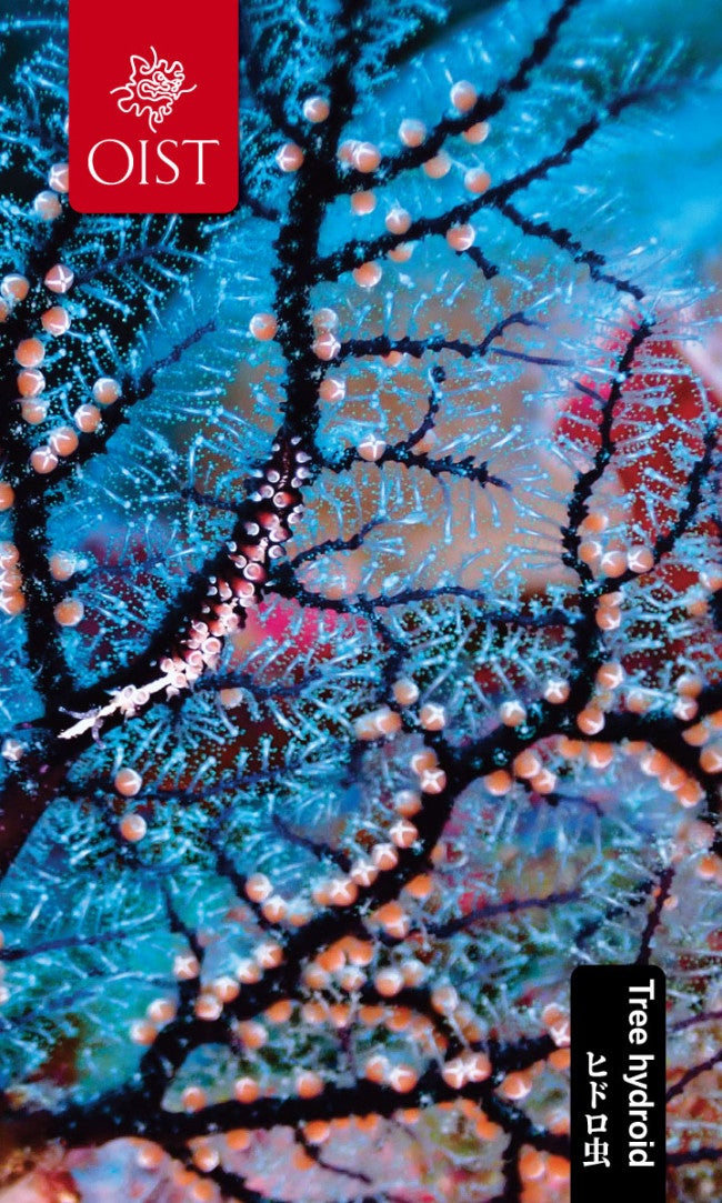
Tree hydroid
Researchers collected this tree hydroid to study the diversity of eukaryotic microbes associated with marine invertebrates. By extracting and analyzing DNA, they identified the hydroid and its microbial community. The black, red and white sea slug, Pleurolidia, visible on one of the branches, feeds preferentially on these hydroids. The orange "fruits" on the tree are specialized reproductive structurescalled gonophores.
Photo: Vera Emelianenko
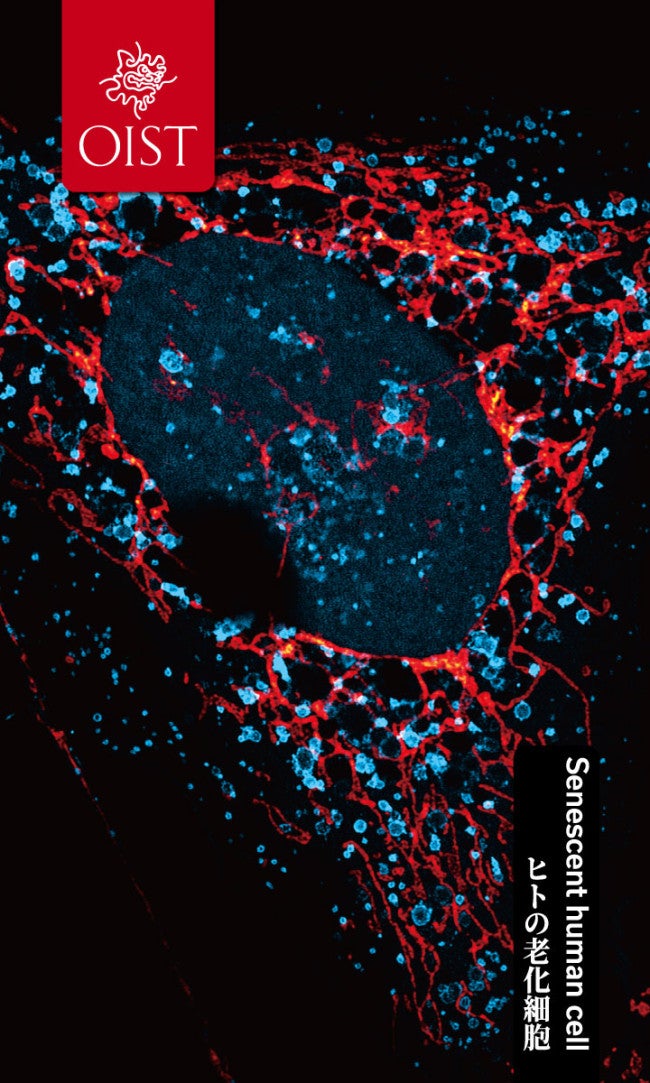
Internal architecture of a senescent human cell
A human cell in senescence, a cellular state linked to stress, damage, and aging. This cell was treated with a compound that selectively targets senescent cells and activates cell death. Using a confocal microscope, mitochondria in red and enlarged vesicles (membrane-bound compartments) in blue can be seen surrounding the central cell nucleus.
Photo: Kamila Kozik
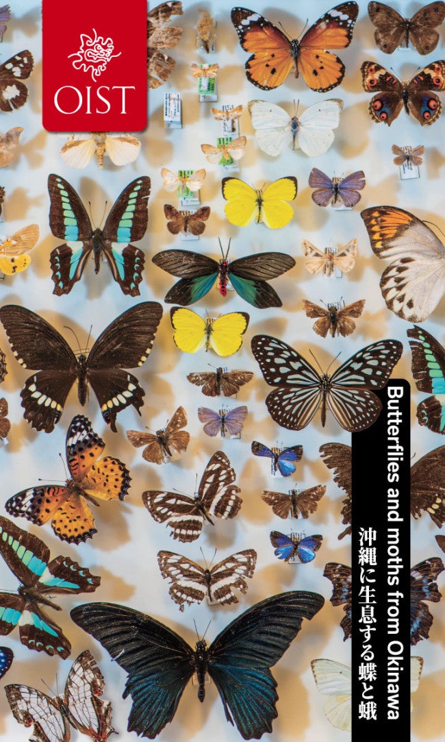
Butterflies and moths from Okinawa
A stunning collection of moths and butterflies from Okinawa that are preserved for environmental research conducted both within and beyond OIST.
Photo: Jeffery Prine, Andrew Scott
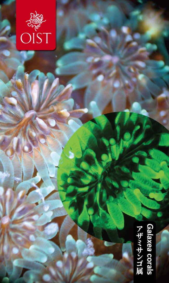
The fluorescent inner lives of Galaxea corals
Corals can become even more colorful when seen under ultraviolet light. The tissues of the stony Galaxea coral, collected locally in Okinawa, are packed with proteins that fluoresce green. The faint red fluorescence is from algae that live inside the coral cells.
Photo: Keenan Guillas
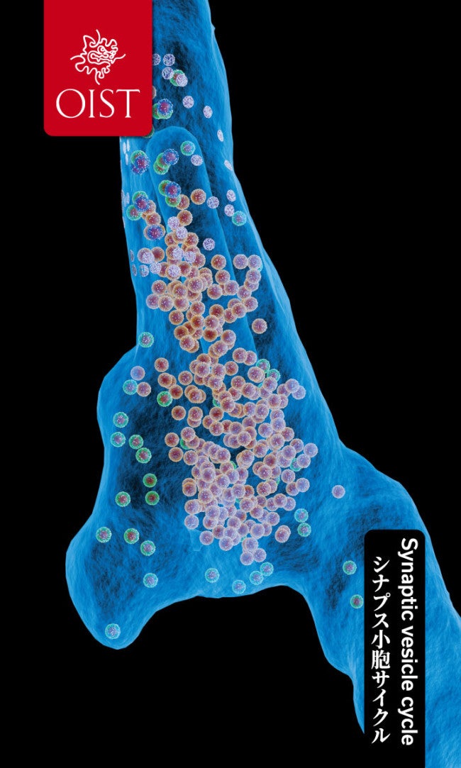
Synaptic vesicle cycle
Researchers use cutting-edge simulations to investigate the synaptic vesicle cycle, one of the fundamental processes that enables neurons in the brain to communicate using chemical signals. Gray spheres represent molecules, while the color of the vesicle indicates its stage in the cycle.
Photo: Iain Hepburn, Jules Lallouette, Andrew Gallimore, Erik De Schutter
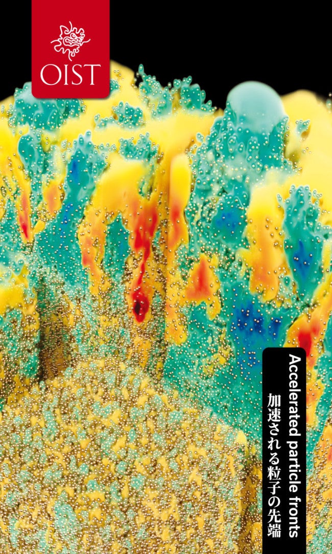
Accelerated particle fronts
Starting at the bottom and proceeding clockwise, this image shows changes that occur over time as a layer of particles immersed in a fluid accelerates upwards. The color represents fluid motion: blue for upwards motion and red for downwards. As the particle rise, turbulent, plume-like structures are generated, enhancing mixing.
Photo: Simone Tandurella, Stefano Musacchio, Guido Boffetta, Marco Edoardo Rosti
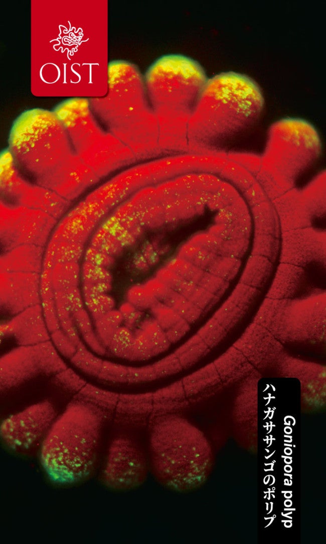
Glowing within: The symbiotic core of a Goniopora polyp
A magnified polyp from the stony coral Goniopora is revealed in three dimensions with vibrant fluorescent color. Polyps —the tiny animals that build coral reefs — depend on a partnership with algae to that live within their cells. Under ultraviolet light, the algae's chlorophyll fluoresces red while the polyp's tissue fluoresces green, highlighting the high density of algae within the coral.
Photo: Sara Hansen
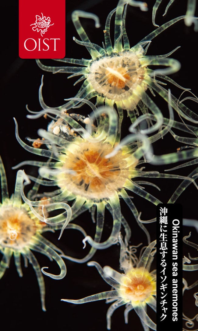
Okinawan sea anemones
In sunlight, Okinawan Aiptasiidae sea anemones readily take up algae, supplementing their diet with products of photosynthesis and turning opaque golden-brown. However, in prolonged darkness, they expel these algae and rely solely on predation, becoming transparent as seen here. Researchers use these anemones as a model organism to study how cnidarians acquire, maintain and lose symbiotic partnerships with algae — a process vital to the health of coral reefs in Okinawa and around the world.
Photo: Jeffery Prine, Andrew Scott
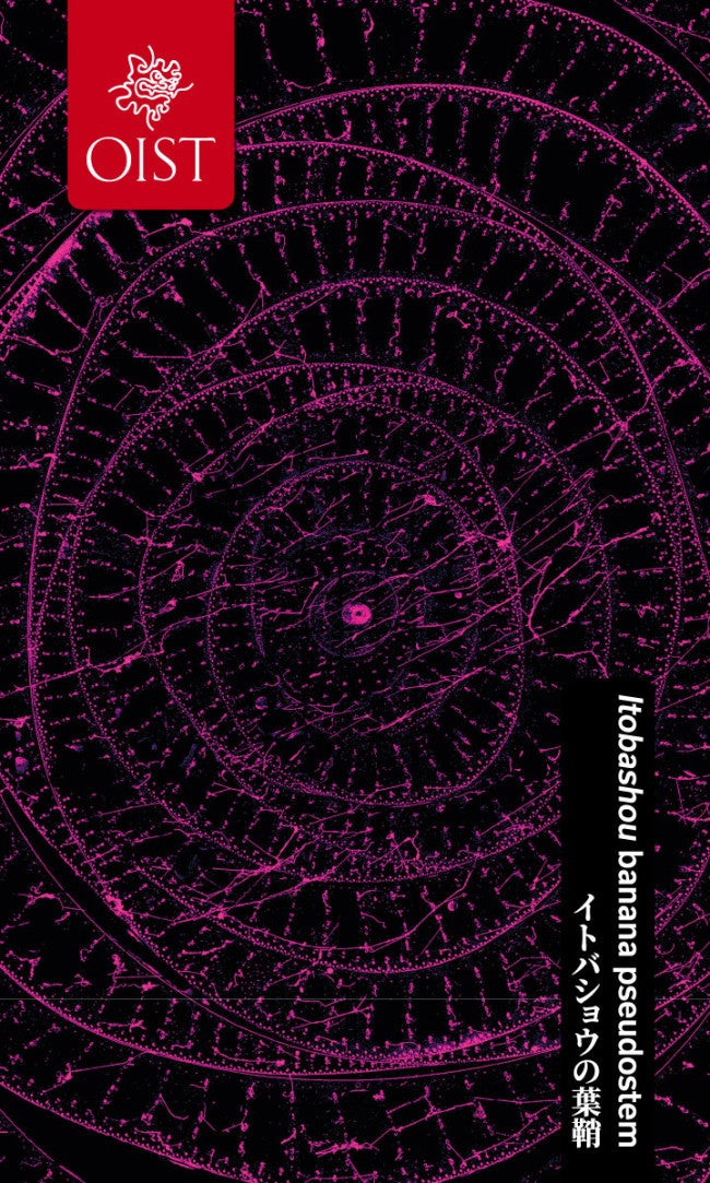
Cross section of Itobashou banana pseudostem
Viewed under a microscope, fibrous tissue in the pseudostem (false stem) of Itobashou, a Ryukyuan banana plant, appears as magenta dots. The false stem is formed from thick, overlapping leaf sheaths. Fibers from the three to four innermost leaf sheaths are extracted and spun into threads, which are used to make Bashofu kimonos, a traditional Ryukuan textile.
Photo: Yoko Nomura, Koji Koizumi
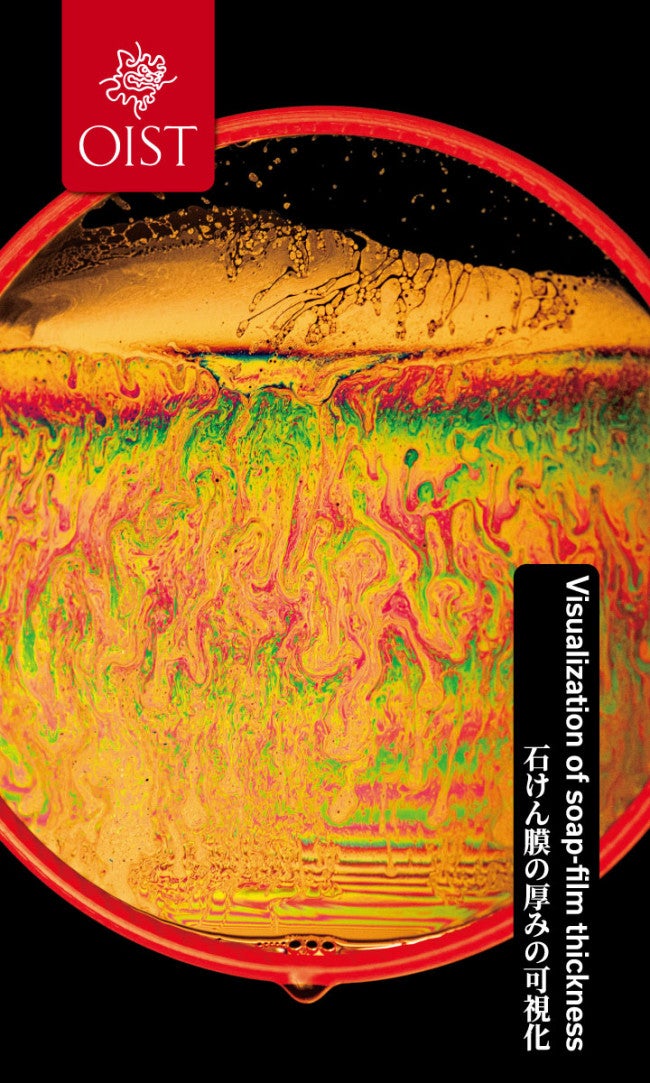
Visualization of soap‑film thickness
Soap film can serve as a valuable tool for exploring fluid behavior and visualizing complex flow structures. Here, the setup demonstrates how variations in a soap film’s thickness can be seen by the way it reflects light. At OIST, similar setups are often used as teaching tools to introduce students and younger audiences to the fundamentals of fluid mechanics, while also opening avenues for future research.
Photo: Jeffery Prine, Andrew Scott
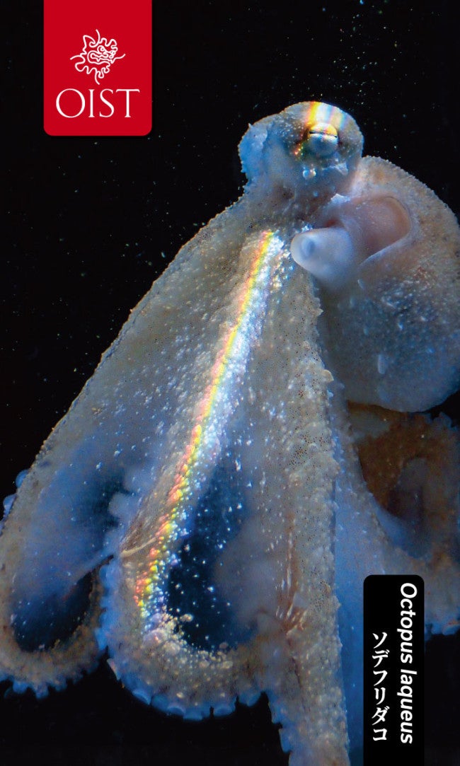
Octopus laqueus
Researchers photographed this octopus, Octopus laqueus,while conducting research on active sleep, a state which resembles human REM sleep. The rainbow colors were created by natural light from the setting sun filtering through other aquarium tanks in the OIST Marine Station.
Photo: Keishu Asada
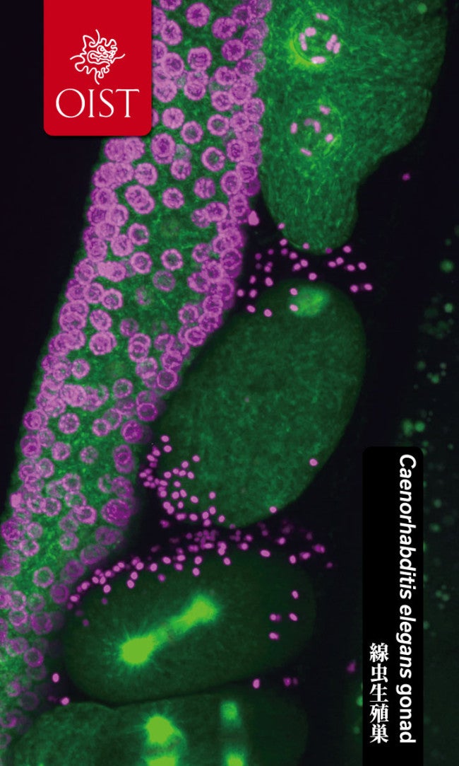
Inheritance
Ultra-fast, live-cell imaging captures the moment a sperm fertilizes an egg in the roundworm, Caenorhabditis elegans, showing DNA in purple and microtubules (the structures that help separate chromosomes during cell division) in green. This research aims to uncover how centrosomes, the main sites where microtubules form, are regulated during development, with implications for understanding genetic disorders, infertility, and cancer.
Photo: Midori Ohta
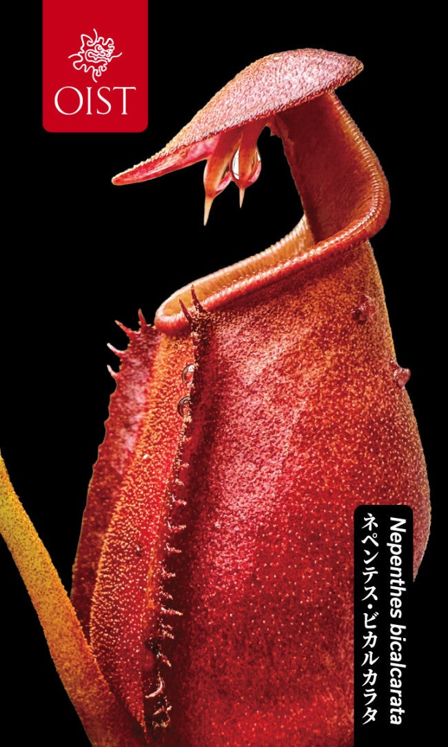
The carnivorous pitcher plant, Nepenthes bicalcarata
A close up of a trap from the carnivorous pitcher plant, Nepenthes bicalcarata, with unique "fang-like" structures that produce nectar to attract and feed ants. The ants, in turn, nourish the plant through their waste, or even by becoming prey.
Photo: David Armitage
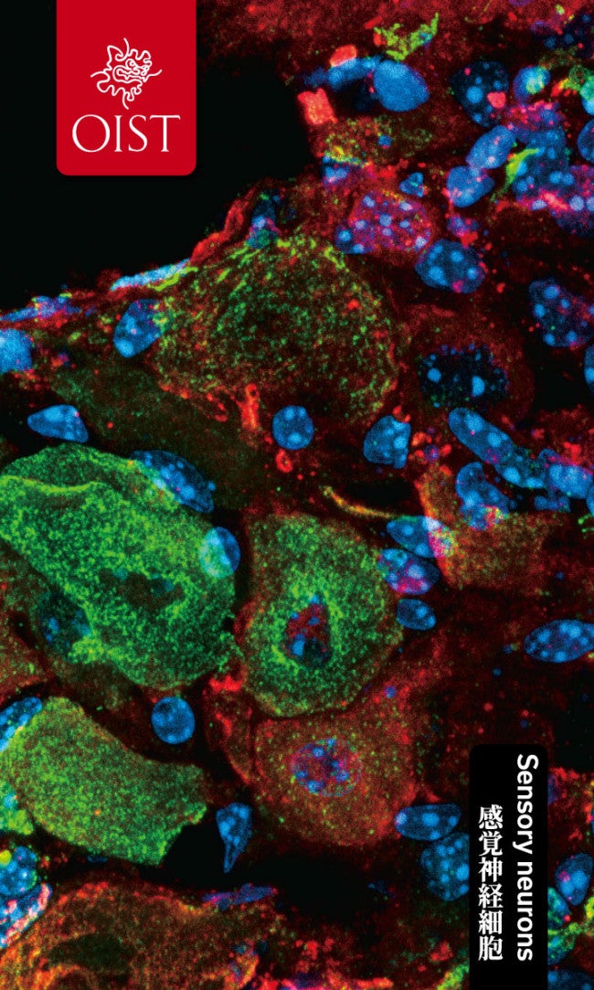
Sensory neurons
An extremely thin section of dorsal root ganglia showing a cluster of sensory neurons located just outside the spinal cord is revealed under a microscope. Different structures within sensory neurons are labeled red and green, while the cell nuclei are stained blue. Researchers use images like this to estimate the number of sensory neurons that survive after sciatic nerve injury, shining light on how neurons recover and regenerate.
Photo: Marco Terenzio
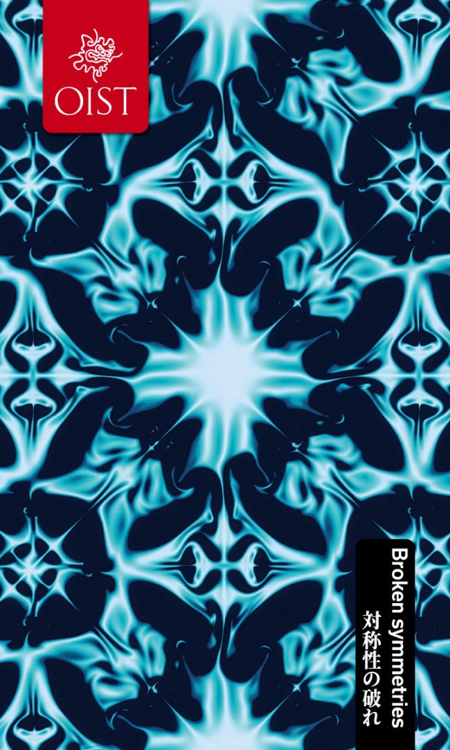
Broken symmetries
Turbulence, the chaotic motion of a fluid, can emerge from simple flows through a series of small, random fluctuations in the fluid that break symmetry. Here, we see a early symmetry-breaking event, one of the first steps in the path towards turbulence.
Photo: Marco Edoardo Rosti
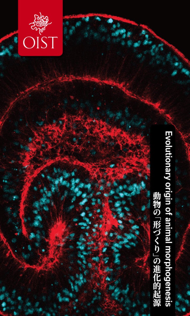
Evolutionary origin of animal body formation
In the embryo of the sea anemone (Nematostella vectensis), which emerged very early in animal evolution, some of its outer cells move inwards to form two cell layers. This process, known as gastrulation, is also observed in most animals, including humans, and is extremely important in the formation of the basic body structure of animals.
Photo: Sen Hadife
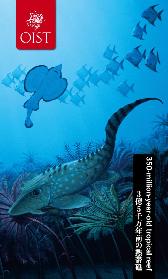
Carboniferous tropical reef
A 350 million-year-old tropical reef from the Carboniferous Period. After a mass extinction 360 million years ago, vertebrates evolved body shapes resembling those of today's reef fishes. However, the dominant species seen here belong to groups now extinct or represented today only by deep-sea relatives, such as ghost sharks.