


Confocal scanning of live zebrafish embryos

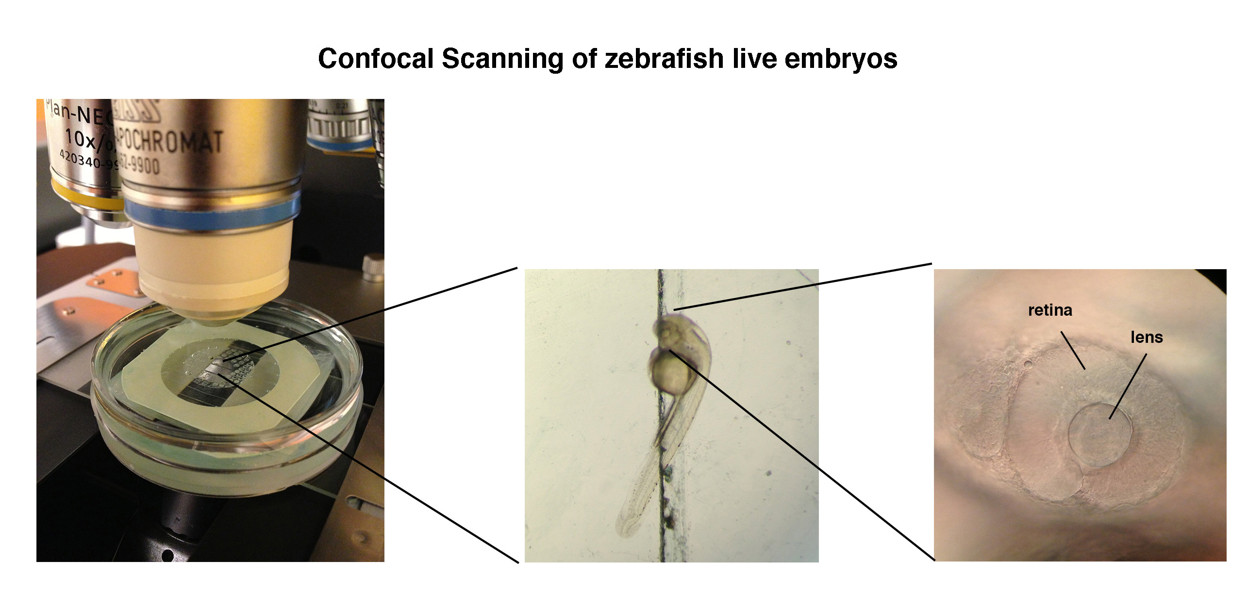
Live zebrafish embryos were immobilized under a confocal microscope so that their eye development could be tracked in real time. The lens of the eye contained bioengineered cells that would express proteins that produced a color visible under the microscope.
Live zebrafish embryos were immobilized under a confocal microscope so that their eye development could be tracked in real time. The lens of the eye contained bioengineered cells that would express proteins that produced a color visible under the microscope.
Date:
14 February 2017
Copyright OIST (Okinawa Institute of Science and Technology Graduate University, 沖縄科学技術大学院大学). Creative Commons Attribution 4.0 International License (CC BY 4.0).
Tags
Research
Share on:


