


29 June 2011
Vesicles, the butlers of the brain
No matter how quickly the delivery truck driver gets a cake to your doorstep, you can't eat it until someone – maybe your butler - opens the door, brings it to the kitchen, and opens the box. Likewise, when most people think about the communication between neurons, they think about electrical and chemical signals passing between neurons like trucks carrying cargo from city to city, house to house. However, there is much more to understand about this process, and scientists at OIST are working to illuminate the minute details of how brain cells called neurons communicate.
We know that inside neurons and the synapses which connect them, vesicles play the role of the household butler, transporting in and out of the cell proteins and other molecules required for proper neuron cell function; however, we do not know a great deal about the behavior of individual vesicles. Dr. Laurent Guillaud of the OIST Cellular & Molecular Synaptic Function Unit (Principal Investigator: Tomoyuki Takahashi) is developing techniques to track single vesicles as they work within the neuron so that he can more fully understand their dynamics and functions.
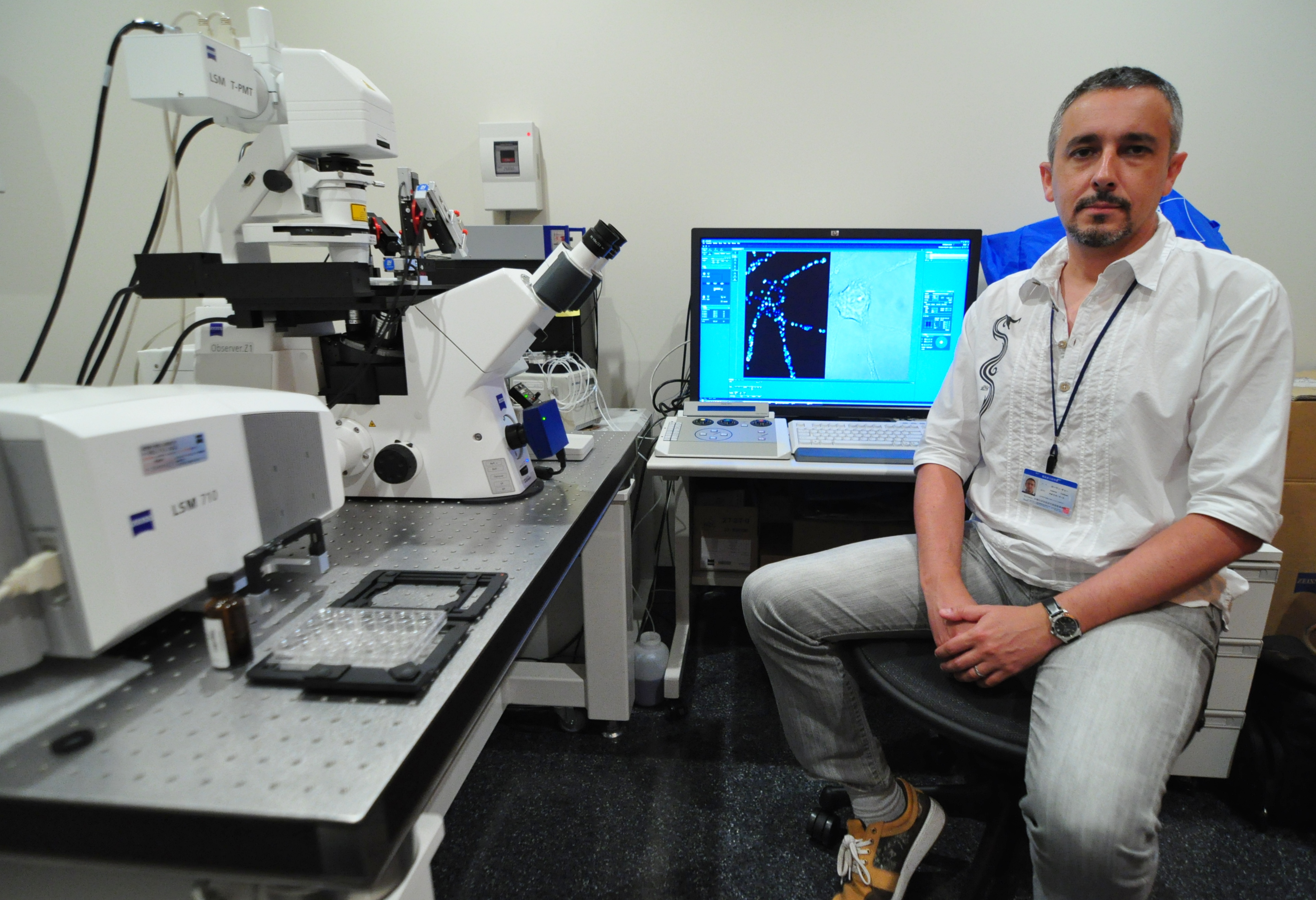
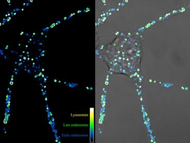
Dr. Guillaud created the accompanying movie using real time imaging confocal microscopy. The vesicles were loaded with a specific pH sensitive fluorescent dye that emits light when excited by the laser of a confocal microscope. He then captured sequential images and processed them to isolate separate views of the cell: one revealing only the light produced by the fluorescent dye; the other, the entire cell. The left panel shows the light produced by the fluorescent dye, and each glowing orb in the left panel corresponds to a vesicle. The center panel of the movie shows the whole cell, and the image in the right panel is a combination of the other two images. This superimposition makes it easy to distinguish the vesicles from the other parts of the neuron.
However, more than just making the vesicles visible, the left panel’s fluorescent light image allows the viewer to distinguish between various types of vesicles. As the pH of the vesicle becomes lower (more acidic), the fluorescent molecules emit more light. By processing the video so that different light intensities are represented by different colors, Dr. Guillaud can monitor the changes in the pH of the vesicles, allowing him to discriminate the different groups of vesicles within the cell.
Vesicles are created, secreted, and reabsorbed as needed. The cell uptakes, or absorbs a molecule by wrapping the cell membrane around it and then detaches this newly-created vesicle from the inner cell membrane in a process called “endocytosis.”
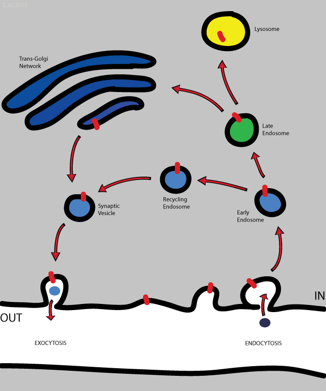
A newly endocytosed vesicle is called an "early endosome" and has a relatively low pH, so it appears blue in the video. As the vesicle's pH becomes lower, it appears green, and this vesicle is referred as a "late endosome." The yellow color in the movie reveals vesicles with the lowest relative pH, which are called "lysosomes.” By color-coding the fluorescence intensity and then superimposing this representation on the neuron image, Dr. Guillaud makes it possible to follow vesicles at these three different stages as they move within the cell.
The movie introduced here and the technique used to create it is one part of Dr. Guillaud's ongoing project to develop methods to accurately track single vesicles as they move within neurons and synapses so that scientists may better understand their role in neural communication.
Just like that cake left on your doorstep, molecules transferred between neurons have to be taken in and out from the cell surface of the neuron and transported for the communication to be complete, so transporting molecules within the cell efficiently is a critical requirement for proper neuronal function. If this traffic is interrupted at any stage, the communication between neurons deteriorates or stops, so more fully understanding the behavior of vesicles could provide new insights into the mechanisms of synaptic transmission – a fundamental aspect of brain function.
For more information, visit the OIST Cellular & Molecular Synaptic Function Unit web page.
Specialty
Research Unit
Submit a press inquiry


