


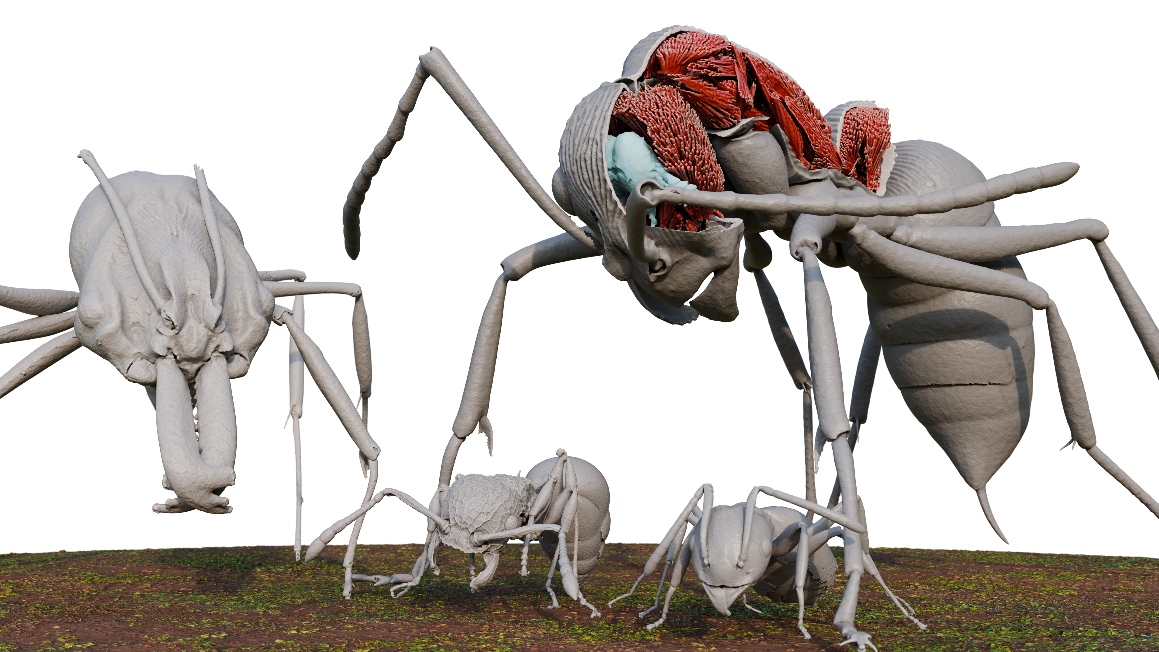
3D renderings made from scans of 4 Okinawan ant species in Antscan

Four Okinawan ants reproduced from Antscan data: Odontomachus kuroiwae (large left), Diacamma cf. indicum (large right), Pristomyrmex punctatus (small left), Technomyrmex brunneus (small right). Diacamma is shown with a portion of its exoskeleton removed, revealing internal organs like part of its nervous system (blue) and muscle fibers (red). The Pristomyrmex and Diacamma specimens were collected by Professor Kazuki Tsuji from the University of the Ryukyus. The other specimens were collected by the OIST OKEON Project’s field team.
Date:
12 February 2026
Credit:
Julian Katzke
Copyright OIST (Okinawa Institute of Science and Technology Graduate University, 沖縄科学技術大学院大学). Creative Commons Attribution 4.0 International License (CC BY 4.0).
Share on:


