


Cryo-EM structure of SVV-ANTXR1 complex

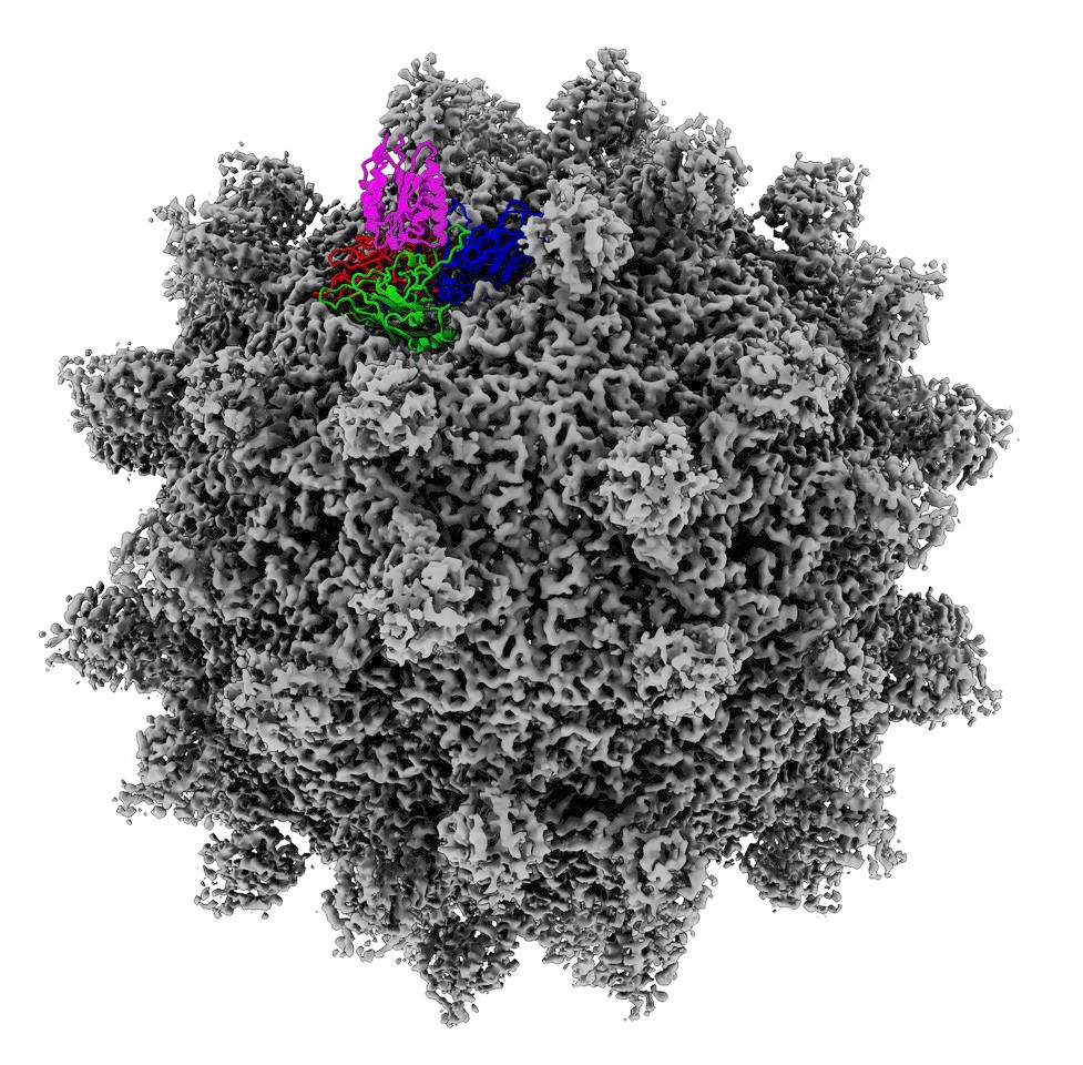
A cryo-EM map of the receptor decorated capsid in which a single protomer was replaced with the atomic model. Seneca Valley Virus capsid proteins are shown in blue, green, and red, and the ANTXR1 receptor is shown in magenta.
A cryo-EM map of the receptor decorated capsid in which a single protomer was replaced with the atomic model. Seneca Valley Virus capsid proteins are shown in blue, green, and red, and the ANTXR1 receptor is shown in magenta.
Date:
30 October 2018
Credit:
OIST Molecular Cryo-Electron Microscopy Unit and University of Otago Centre for Electron Microscopy
Copyright OIST (Okinawa Institute of Science and Technology Graduate University, 沖縄科学技術大学院大学). Creative Commons Attribution 4.0 International License (CC BY 4.0).
Tags
Research
Share on:


