


31 July 2019
New Approach Developed for Brain Imaging Studies

Scientific discoveries are exciting, but scientists must also develop new approaches to make such discoveries possible. Researchers at the Okinawa Institute of Science and Technology Graduate University (OIST) have written a review that summarizes an imaging method using voltage to measure brain activity. The approach allows scientists to probe central questions in brain research, including how brain activity might give rise to behavior.
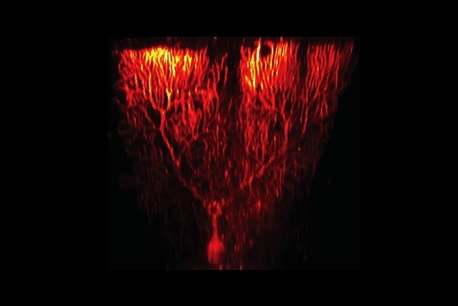
Writing in Frontiers of Cellular Neuroscience, the team distils 20 years of research into a primer on their “voltage imaging” approach, a diverse set of methods that use dyes and microscopes to visualize brain activity. This can be used to study individual neurons and entire sections of the brain, in both tissue cultures and awake animals.
The approach is especially powerful because it can quickly capture changes in brain activity — if necessary, on the scale of nanoseconds. It can be combined with other imaging techniques, and the dyes can safely persist in the brain for several weeks, opening the possibility for more complex, extended studies.
“We are pleased to finally share this summary of our voltage imaging approach,” says Professor Bernd Kuhn, who wrote the primer with postdoctoral scholar Dr. Christopher Roome. “The upcoming chapter in Multi-Photon Microscopy by Springer Nature Neuromethods, meanwhile, will provide experimental protocols.”
Voltage imaging 101
To image the brain in action, researchers first inject a fluorescent dye into either tissue or a single cell. The glow of these dye molecules is eventually detected by light sensors. To deliver the dye into a cell, a researcher quickly jolts the membrane with electricity. This momentarily opens the membrane so that the dye molecules can pass through. Once in place, the dye can get to work.
The fluorescent dye molecules constantly emit light. When a brain cell is activated, an impulse moves along the cell — which changes the dye’s electronic structure and the intensity of light it emits. Scientists use sensors to detect this change, and from these data they draw conclusions about what is happening in the brain.
The approach has already generated interesting results. Last year, Kuhn’s team visualized, for the first time, how particular neurons in an awake mouse are activated. This finding illuminated a key step in how brain cells process information and suggested how behaviors might emerge as a result.
But despite successes, the method has limits. In particular, injecting dye molecules into neurons is a difficult task. Looking down a microscope, a researcher must properly place a probe beside the cell membrane before delivering a shot of electricity and injecting the dye with just the right force. The procedure often misfires, misplacing dye molecules and damaging cells.

Kuhn plans to refine the approach so that it no longer relies on human dexterity. As well as being frustrating, this manual activity slows down the process. Finding a new way of delivering the dyes will allow researchers to work much faster.
Until then, however, Kuhn remains confident that this voltage imaging method will be useful to researchers asking tough questions about the brain.
“Neuroscience often asks how complex behaviors emerge from activity in the brain,” he says. “To answer such questions, we need to be able to study brain activity at many levels — our approach is a new tool to do just that.”
Following the recent primer in Frontiers of Cellular Neuroscience, the OIST team’s work will form the basis of a chapter in the forthcoming Multi-Photon Microscopy book by Springer Nature Neuromethods — out August 2019.

Specialties
Research Unit
Submit a press inquiry


