


Filling a Purkinje neuron with fluorescent dye

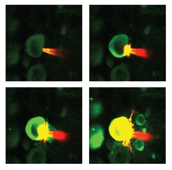
Looking down a microscope, researchers place a probe beside the cell membrane of a Purkinje neuron and inject fluorescent dye. Once correctly in place, the dye molecules provide insight on the brain cell’s activity.
Looking down a microscope, researchers place a probe beside the cell membrane of a Purkinje neuron and inject fluorescent dye. Once correctly in place, the dye molecules provide insight on the brain cell’s activity.
Date:
01 August 2019
Copyright OIST (Okinawa Institute of Science and Technology Graduate University, 沖縄科学技術大学院大学). Creative Commons Attribution 4.0 International License (CC BY 4.0).
Tags
Research
Share on:


