


SEM and STEM mode versus electron holographic imaging mode

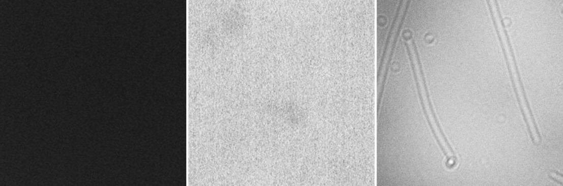
The two conventional modes of a scanning electron microscope (SEM and STEM; left and center) were unable to generate images of the biomolecules. However, holographic imaging mode (right), can be used to image biomolecules, such as the tobacco mosaic virus shown.
The two conventional modes of a scanning electron microscope (SEM and STEM; left and center) were unable to generate images of the biomolecules. However, holographic imaging mode (right), can be used to image biomolecules, such as the tobacco mosaic virus shown.
Date:
16 March 2020
Credit:
Modified from M Cheung, H Adaniya, C Cassidy, M Yamashita, T Shintake. Low-energy in-line electron holographic imaging of vitreous ice-embedded small biomolecules using a modified scanning electron microscope. Ultramicroscopy, 209 (2020) 112883, Fig 3.
Copyright OIST (Okinawa Institute of Science and Technology Graduate University, 沖縄科学技術大学院大学). Creative Commons Attribution 4.0 International License (CC BY 4.0).
Tags
Research
Share on:


