


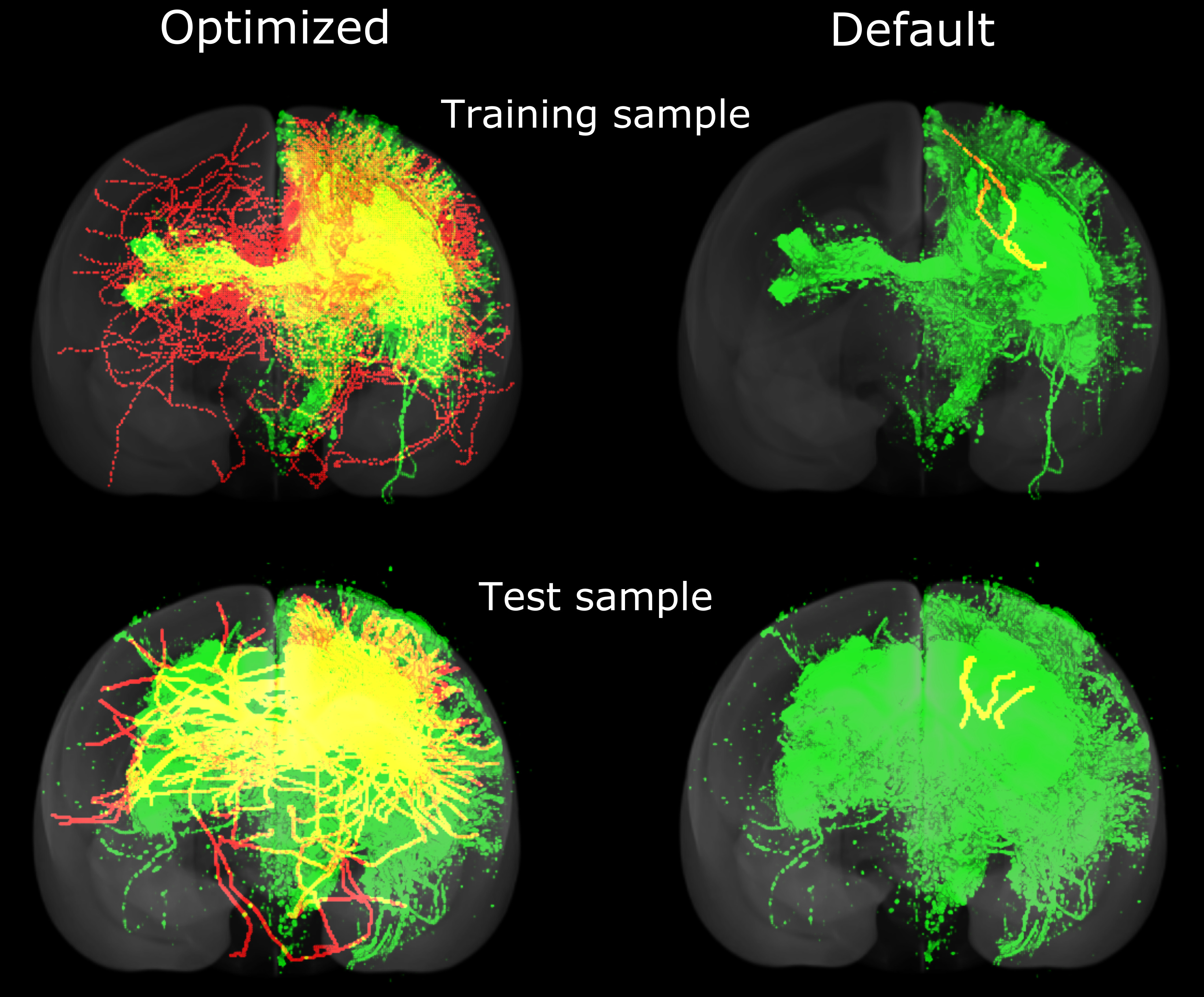
Comparing optimized and default algorithms on training brains and test brains

The green represents nerve fibers detected by injecting a fluorescent tracer at a single point. The red represents nerve fibers detected using a diffusion MRI-based fiber tracking algorithm. Only the nerve fibers that also connected up to the point where the tracer was injected are shown. The yellow represents nerve fibers that were detected using both techniques. The results show that the optimized algorithm performed better than the default algorithm, not only on a brain it was trained on, but on a previously unseen brain. The optimized algorithm detected a higher number of fibers and also fibers that stretched longer distances.
The green represents nerve fibers detected by injecting a fluorescent tracer at a single point. The red represents nerve fibers detected using a diffusion MRI-based fiber tracking algorithm. Only the nerve fibers that also connected up to the point where the tracer was injected are shown. The yellow represents nerve fibers that were detected using both techniques. The results show that the optimized algorithm performed better than the default algorithm, not only on a brain it was trained on, but on a previously unseen brain. The optimized algorithm detected a higher number of fibers and also fibers that stretched longer distances.
Copyright OIST (Okinawa Institute of Science and Technology Graduate University, 沖縄科学技術大学院大学). Creative Commons Attribution 4.0 International License (CC BY 4.0).
Tags


