


22 March 2022
Staying alive: Scientists eye up gene required for the survival of an important retinal neuron

Highlights
- Retinal ganglion cells are a type of neuron found in the eye that are essential for vision
- A new study on zebrafish has found that a protein called Strip1 is required to keep these cells alive during and after development
- When Strip1 is absent within retinal ganglion cells, a protein called Jun is activated, which ultimately triggers the cells to die
- The death of retinal ganglion cells leads to developmental defects in the structure of the retina, within a region called the inner plexiform layer
- Uncovering the role of Strip1 could lead to promising new research avenues for treating glaucoma – one of the leading causes of blindness that occurs when retinal ganglion cells die
Press Release
Researchers at the Okinawa Institute of Science and Technology Graduate University (OIST) in Japan have identified a gene necessary for the survival of retinal ganglion cells – a class of neurons located in the retina that are critical for vision.
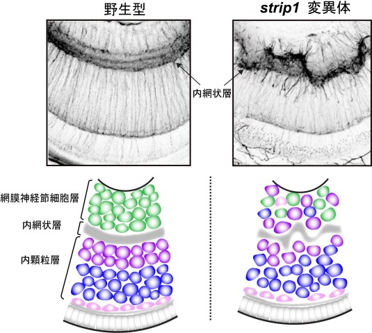
Reporting in elife on March 22, 2022, the scientists found that when zebrafish embryos developed without a working version of a gene called strip1, most retinal ganglion cells died, and inner retinal layers formed abnormally. As well as providing fundamental insights into how the retina develops, discovering how the protein encoded by the strip1 gene keeps retinal ganglion cells alive also opens promising new avenues into treating diseases like glaucoma.
“Glaucoma is one of the leading causes of blindness worldwide, with the loss of vision caused by the death of retinal ganglion cells,” said Professor Ichiro Masai, head of the Developmental Neurobiology Unit at OIST. “In the future, uncovering the full Strip1 protein pathway inside retinal ganglion cells could help scientists find ways to slow down, or even prevent, their death in patients with glaucoma.”
Prof. Masai, along with his PhD student, Mai Ahmed, first became interested in the strip1 gene due to its role in correctly forming inner retinal layers.
“When you look at the retina under a microscope, you can see that it has this beautifully organized structure. Different neurons are stacked on top of each other, with synaptic layers in between where the neurons connect and communicate to each other,” said first author, Mai Ahmed. “These neurons carry electrical signals from the photoreceptors that detect light all the way to the visual centers of the brain. Without proper wiring of these retinal circuits, vision is compromised.”
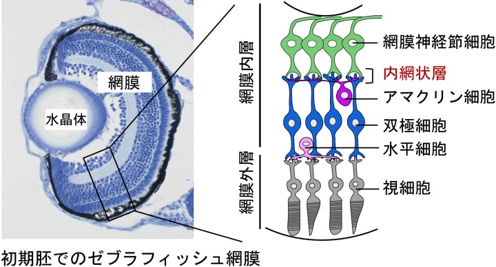
Back in the early 2000s, scientists at RIKEN in Japan, including Prof. Masai, created hundreds of zebrafish embryos that contained random mutations. They saw that one of these mutations caused a defect in a synaptic layer in the retina, known as the inner plexiform layer. Further research at OIST identified that the mutation occurred within the gene, strip1.
Seeking to uncover the reason behind this retinal defect, Prof Masai’s unit labelled the three different types of neurons that connect in the inner plexiform layer – retinal ganglion cells, amacrine cells and bipolar cells – to see how they developed when strip1 was mutated.
Surprisingly, the researchers found that while all three neuron types developed an incorrect form and position, only the retinal ganglion cells died. As these cells died, the other neuron types replaced them, leading to a disordered layer. The Strip1 protein is therefore essential for keeping ganglion cells alive, and thus maintaining the correct form of the inner plexiform layer.
When the researchers looked at the underlying mechanism, they found that Strip1 suppresses the activity of Jun, a protein associated with cell death. Jun, and the pathway it acts within, is triggered when neurons are under stress. If enough of the Jun protein is activated, the cell dies.
“This process of cell death, called apoptosis, is a fail-safe mechanism for removing stressed cells that can’t be repaired,” explained Prof. Masai. “But we believe retinal ganglion cells are placed under immense metabolic stress during normal development, when they form the incredibly long optic nerve. So Strip1 is needed inside retinal ganglion cells to protect them from the Jun apoptotic pathway.”
Importantly, Jun, and the pathway it acts within, is known to play a role in the death of retinal ganglion cells in patients with glaucoma.
“Glaucoma typically occurs when pressure damages the optic nerve, placing the retinal ganglion cells under immense stress, and activating Jun,” said Mai. “If, in future research, it’s found that Strip1 can suppress Jun in mammalian glaucoma models too, this would open the door to many new therapies.”

Article Information
Title: Strip1 regulates retinal ganglion cell survival by suppressing Jun-mediated apoptosis to promote retinal neural circuit formation
Journal: elife
Authors: Mai Ahmed, Yutaka Kojima, and Ichiro Masai
DOI: 10.7554/eLife.74650
Date:2022.03.22
Specialties
Research Unit
Submit a press inquiry


