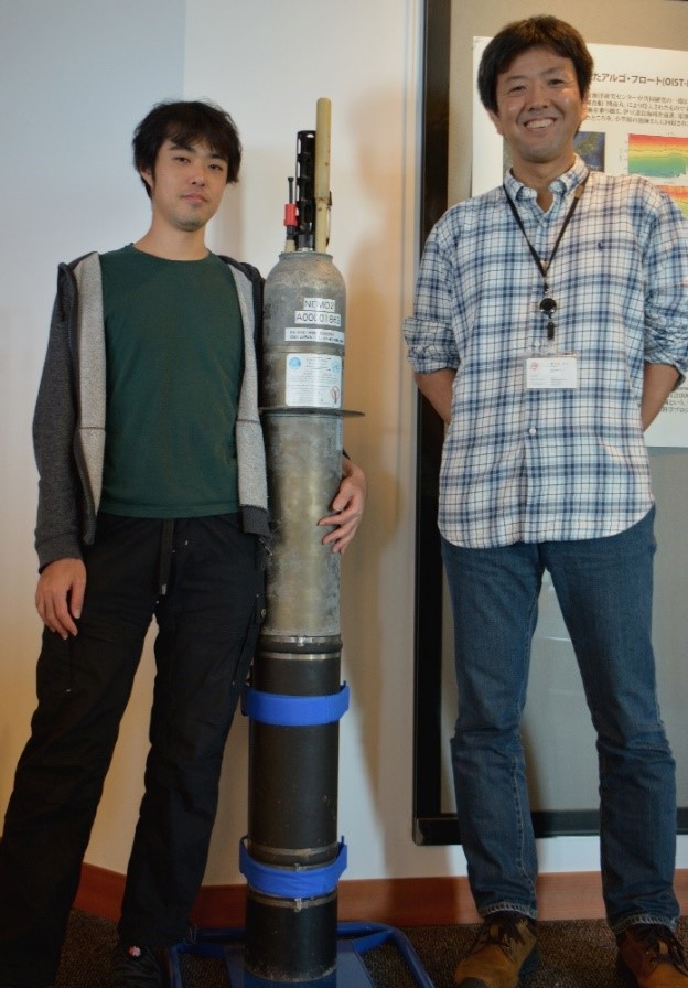
Post-doctoral scholar, Yuichi Nakajima and Prof. Satoshi Mitarai (right) of the Marine Biophysics Unit at OIST Nakajima and Mitarai (right) pictured with a deep-ocean profiling float. They deployed 10 of these floats to track potential larvae dispersal from hydrothermal vents. Date: 18 March 2016 Copyright OIST (Okinawa Institute of Science and Technology Graduate University, 沖縄科学技術大学院大学). Creative Commons Attribution 4.0 International License (CC BY 4.0). Download full-resolution image Share on: Related Images Principle Idea of PhysioDesigner To simulate multi-level physiological responses it is necessary to understand how physiological phenomena occurring at one level influence or govern other levels. Leptoceratops Artistic rendering of Leptoceratops, a likely relative of Serendipaceratops arthurcclarkei by Peter Trusler. X-ray micro-CT X-ray micro-CT 3D model of ant Burriel-Ha’s 3D model of the ant head and first segment next to the 3D image on the computer used to print it. Museum fly specimen Comparison of one of the fly specimens used in the study before and after DNA extraction. There is minimal damage to the eye and the wings, but the specimen is mostly unharmed from the new extraction technique.
Principle Idea of PhysioDesigner To simulate multi-level physiological responses it is necessary to understand how physiological phenomena occurring at one level influence or govern other levels.
Leptoceratops Artistic rendering of Leptoceratops, a likely relative of Serendipaceratops arthurcclarkei by Peter Trusler.
3D model of ant Burriel-Ha’s 3D model of the ant head and first segment next to the 3D image on the computer used to print it.
Museum fly specimen Comparison of one of the fly specimens used in the study before and after DNA extraction. There is minimal damage to the eye and the wings, but the specimen is mostly unharmed from the new extraction technique.