


Diagram of the zebrafish eye

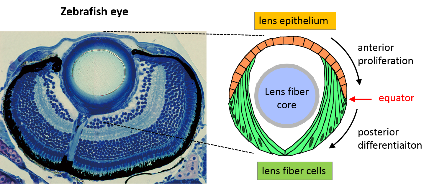
Diagram of the zebrafish eye. Left: photograph of the zebrafish eye under a microscope, with the anterior region situated at the top of the photograph and the posterior region at the bottom. Right: diagram of the zebrafish eye lens depicting where the lens epithelial and fiber cells are relative to the rest of the eye.
Diagram of the zebrafish eye. Left: photograph of the zebrafish eye under a microscope, with the anterior region situated at the top of the photograph and the posterior region at the bottom. Right: diagram of the zebrafish eye lens depicting where the lens epithelial and fiber cells are relative to the rest of the eye.
Copyright OIST (Okinawa Institute of Science and Technology Graduate University, 沖縄科学技術大学院大学). Creative Commons Attribution 4.0 International License (CC BY 4.0).


