


Analysis of zebrafish lens growth
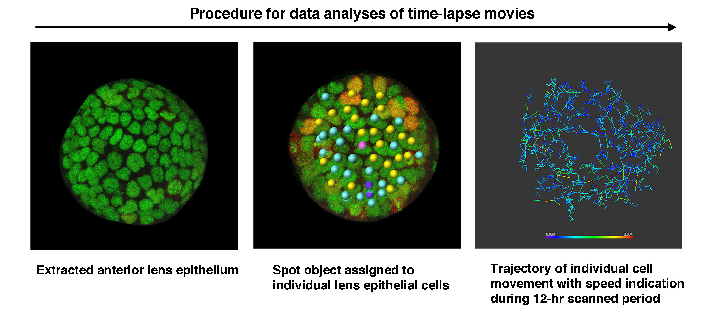
Tracking procedure of individual cell growth. Left: Individual lens epithelial cells are observed using a confocal microscope. Middle: Each cell is assigned a spot of a specific color, depending on the cell’s classification: dividing, non-dividing, or dead/dying. Right: The movement patterns of each cell are tracked and marked by a trajectory line. The color of the line indicates the speed at a particular location.
Tracking procedure of individual cell growth. Left: Individual lens epithelial cells are observed using a confocal microscope. Middle: Each cell is assigned a spot of a specific color, depending on the cell’s classification: dividing, non-dividing, or dead/dying. Right: The movement patterns of each cell are tracked and marked by a trajectory line. The color of the line indicates the speed at a particular location.
Copyright OIST (Okinawa Institute of Science and Technology Graduate University, 沖縄科学技術大学院大学). Creative Commons Attribution 4.0 International License (CC BY 4.0).


