24 May 2024
Lighting up the brain: What happens when our ‘serotonin center’ is triggered?

Our brains are made of tens of billions of nerve cells called neurons. These cells communicate with each other through biomolecules called neurotransmitters. Serotonin, a type of neurotransmitter, is produced by serotonin neurons in our brains and influences many of our behavioral and cognitive functions such as memory, sleep, and mood.
Using mice, scientists at the Okinawa Institute of Science and Technology (OIST) and their collaborators from Keio University School of Medicine have studied the main source of serotonin in the brain – the dorsal raphe nucleus (DRN). By studying how activating the brain’s ‘serotonin center’ affects awake animals for the first time, they found that serotonin from the DRN activates brain areas that affect behavior and motivation.
“Learning about the brain’s serotonin system can help us understand how we adapt our behaviors and how mood therapy medication works. But it was hard to study how serotonin from the DRN affects the entire brain. First, because electric stimulation of the DRN can also activate neurons that don’t use serotonin to communicate with each other, and second, using drugs can affect other serotonin in the brain,” explained Dr. Hiroaki Hamada, a former PhD student at OIST’s Neural Computation Unit and lead author of a paper on this study published in the journal Nature Communications.
Previous studies by researchers at the Neural Computation Unit have shown that serotonin neurons in the DRN promote adaptive behaviors in mice associated with future rewards. Dr. Hamada and his collaborators wanted to understand the mechanisms in the brain that cause these adaptive behaviors.
“We knew that DRN serotonin activation has strong effects on behavior, but we didn’t know how this serotonin activation affects different parts of the brain,” Prof. Kenji Doya, leader of the Neural Computation Unit, stated.
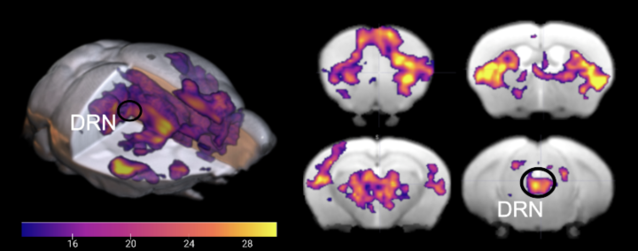
Observing the entire brain’s response to DRN serotonin activation
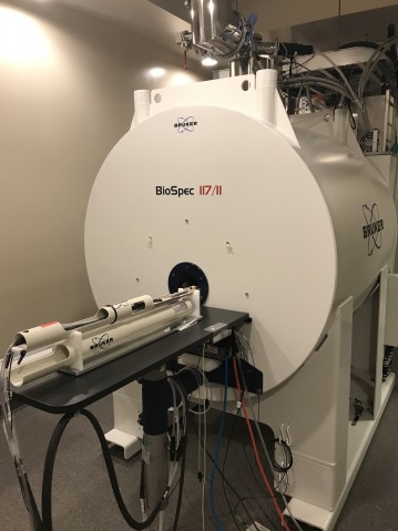
The researchers used a novel technique called opto-functional MRI to address this question. They used a method called optogenetics to selectively activate serotonin neurons in the DRN with light and observed the entire brain’s response using functional MRI (Magnetic Resonance Imaging). They utilized the latest MRI scanner with a strong magnetic field to achieve the high resolution needed to study the small brains of mice. The mice were put in the MRI scanner and serotonin neurons were stimulated at regular intervals to see how this affected the whole brain.
They found that DRN serotonin stimulation causes activation of the cerebral cortex and the basal ganglia, brain areas involved in many cognitive functions. This result was very different from a previous study performed under anesthesia. Additionally, the brain's response to serotonin stimulation is strongly linked to the distribution of serotonin receptors (proteins activated by serotonin) and the connection patterns of DRN serotonin neurons.
“We clearly see from the high-field MRI images which areas in the brain are activated and deactivated during the awake state and under anesthesia when we activate serotonin neurons in the DRN,” Dr. Hamada said. “A previous study showed that the cerebral cortex and the basal ganglia were mostly deactivated under anesthesia, which we also observed, however, in awake states these areas are significantly activated.”
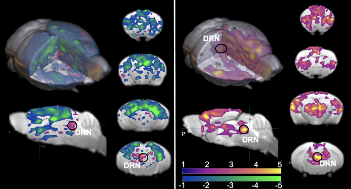
The cerebral cortex and the basal ganglia are parts of the brain critical for many cognitive processes, including motor activity and behaviors to gain rewards such as food and water. Activation of DNR serotonin neurons can therefore lead to changes in motivation and behavior.
Patience and stimulating your own serotonin
Combining the new technique of high field MRI and optogenetics presented many obstacles that Dr. Hamada had to overcome. “We introduced and adapted a method previously used by our collaborators and established many new procedures at OIST. For me, the main challenge was using the new MRI machine at the time, so I needed to have patience and stimulate my own serotonin. I started doing a lot of exercise after that,” he laughed.
Seeing activations in the DRN for the first time was a standout moment for Dr. Hamada. In the beginning, he used the same light intensity that his collaborators used, but this was too weak to see the brain responses in the MRI. He then used bigger optical fibers and increased the intensity to stimulate the DRNs.
Prof. Doya noted that the next important milestone to achieve is understanding exactly how this brain-wide activation of serotonin occurs: “It’s important to find out what is the actual molecular mechanism allowing this activation in our brain. People who would like to get better at adjusting their behavior and thinking in different situations could also find it helpful to learn more about how serotonin helps control our moods.”
To read more about research on the effects of serotonin from Prof. Doya’s unit, please see here.
Article Information
Research Unit
Submit a press inquiry