


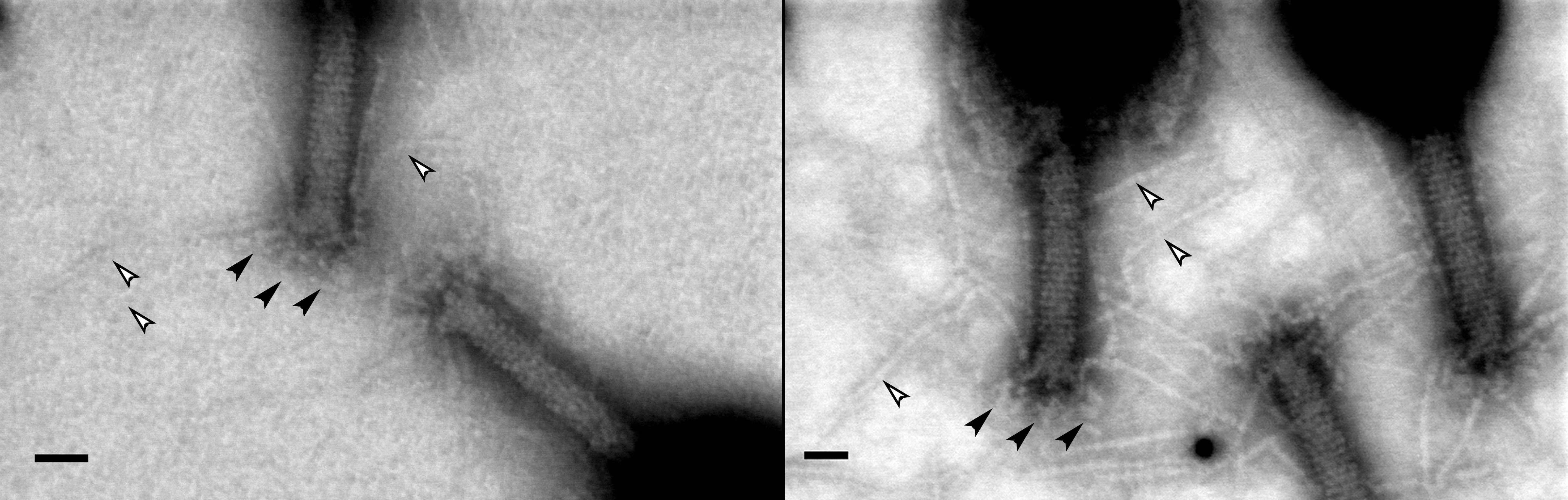
Using a graphene film provides a much high contrast revealing details.

Electron Microscopy images of bacteriophage T4 viruses. The left image was made using conventional carbon film, the right image using the single atom thin graphene layer. More details about the tail and the limbs of the viruses can be discerned. The black bar used for scale is 20 nanometers (0.00000002 meters).
Date:
09 June 2017
Copyright OIST (Okinawa Institute of Science and Technology Graduate University, 沖縄科学技術大学院大学). Creative Commons Attribution 4.0 International License (CC BY 4.0).
Tags
Research
Share on:


