


22 September 2025
J-PEAKS Funding Elevates Core Facilities and Advances Cryo-EM Research

OIST has been awarded ¥5.5 billion over five years through MEXT’s J-PEAKS program. A key focus of the award is enhancing Core Facilities, which underpins research across OIST by providing state-of-the-art equipment and expert technical support.
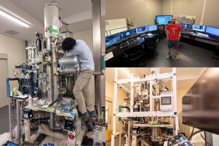
Using J-PEAKS funding, Core Facilities recently purchased two new microscopes for cryoelectron imaging*. Both are from the Japanese producer JEOL and are considered top of the line.
Oleg Sitsel, whose lab, Marine Structural Biology Unit, studies marine organisms using cryoelectron microscopy, notes that the machines, CRYO ARM 200 II and CRYO ARM 300 II, are equipped with:
- Cold field emission guns, which produce very bright electron beams with nearly no energy spread,
- In-column energy filters, which improve the signal-to-noise ratio of the acquired images, and
- State of the art detectors for acquiring those images.
- The CRYO ARM 300 II is also equipped with a unique high-resolution polepiece configuration.
“Taken together, this will enable us to acquire high-quality images of proteins for determining their structures at near-atomic resolution,” says Sitsel. “High-resolution protein structures help us explain how these biological nanomachines function and inform us of ways how we can tweak them to serve our needs if necessary. Also, seeing how proteins and other intracellular components are arranged within cells helps us understand how cells work with unprecedented detail.”
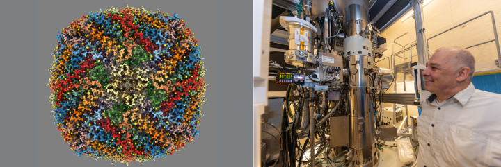
Professor Matthias Wolf, who leads the Molecular Cryo-Electron Microscopy Unit, agrees. “This may be the world's best cryo-transmission electron microscope for single particle analysis,” says Wolf. “We have already reached the current resolution record in cryo-EM — with this scope, we will be able to break it. I hope to visualize hydrogen atoms, which will have a big impact on enzymology and drug design.”
OIST will continue to leverage support from the J-PEAKS initiative to enhance its core facilities with cutting-edge research equipment, building a robust foundation that empowers researchers to pursue bold scientific challenges.
*While the two microscopes were funded through separate sources, support from the J-PEAKS initiative enabled a strategic, simultaneous procurement—facilitating favorable negotiations and the acquisition of high-performance equipment.


