


14 February 2024
Deciphering the mysteries of cell division

The human body consists of millions of cells, many of them dividing every day to keep our tissues healthy and functional. The process in which cells divide is complex and things can go wrong at any stage causing severe diseases, including cancer. Studying these cellular processes is the goal of Dr. Franz Meitinger. Together with Dr. Midori Ota, who is a Science and Technology Associate, they established the Cell Proliferation and Gene Editing Unit at OIST in 2022.
The goal of the unit is to better understand cell division and how individual cells recognize defects that occur during this process. But, trying to look both ways, the lab also investigates what happens after something goes wrong and searches for potential drug targets in cancer cells.
Studying these mechanisms, which take place deep within a cell, can be challenging and relies on several molecular biological methods. Researchers in the unit use model-cell lines, which are populations of individual cells that are kept alive in cultures, to understand the inner workings of cells. While some of them are healthy, other lines originate from samples of tumors, and therefore carry cancer specific genetic mutations. By editing genes and microscopic imaging of live cells, the researchers can observe processes inside the cells in real time.
First, Dr. Meitinger had other career ambitions than going into science. “I grew up on a farm in a small village in Bavaria,” says Dr. Meitinger, who got his first job at the age of sixteen. After leaving school, he worked as a technician in a chemical manufacturing company for a few years but driven by his strong curiosity to understand how the world works, he decided to go back to school and eventually studied biology at university.
After obtaining his Ph.D. at the German Cancer Research Center in Heidelberg, he continued his work on cell division as a postdoc at the Ludwig Institute for Cancer Research in San Diego. There he discovered the mitotic stopwatch mechanism, which uses the time it takes for a cell to go through mitosis to detect defects in cell division.
If a mistake occurs during cell division the process normally stops until the problem is fixed. In rare cases, the defective cells cannot correct the mistake and continue to move forward with cell division thereby creating an irreversible damage. When comparing the time it takes for healthy and faulty cells to divide, it becomes apparent that the latter divide at a much slower pace. The cells themselves can sense this time difference and upon discovering that it divides too slowly, specialized proteins are activated. This mechanism is called the mitotic stopwatch pathway and helps to remove damaged cells from the proliferating pool.
There at OIST, the Meitinger unit is continuing this research focusing on cell division. “We are interested in the mechanisms that can sense when something goes wrong,” emphasizes Dr. Meitinger. Comparing cultured cancerous and healthy cells allows the lab to go a step further, using methods such as genome wide screens they also work on identifying cancer specific changes that could be used as treatment targets. “I want to help improve the world, which is a long way if you think about basic biology,” says Dr. Meitinger. But cell division still holds many mysteries which are waiting for researchers to uncover them.
As a Science and Technology Associate, Dr. Ota independently works on understanding the regulation of centrosomes, which segregate chromosomes during cell division. In collaboration with the Meitinger unit, she uses human cell lines and Caenorhabditis elegans. The latter is a translucent roundworm commonly used as a model system and was originally established by Dr. Sydney Brenner, the first President of the Okinawa Institute of Science and Technology Promotion Corporation.
For Dr. Ota, it was her passion for dance that eventually led to science. “I wanted to be a ballerina,” she says. “When I was dancing, at the age of fifteen, I went to a sports science lecture,” Dr. Ota remembers. This talk proved to be very helpful, as the anatomical knowledge presented there helped the young dancer to reduce pain during training. Experiencing first-hand how science can improve wellbeing, she decided to pursue a career in science herself.
During her time as a high school student, she became fascinated by how cells, despite possessing identical genetic information, end up with different functions and identities. “You know, whenever people have to study for exams, they procrastinate,” reminisces Dr. Ota whose preferred method of procrastination during study for exams was shaping her eyebrows.
“A few weeks after removing a hair, at the same place, the same hair had grown back. That got me interested in what gives a cell a specific identity”. This initial interest in how cells work led her to study centrosomes, which are complex protein structures serving as nucleators for thin tube-like structures, called microtubules. The centrosomes and microtubules are essential during cell division as they divide the DNA equally between the two newly forming daughter cells.
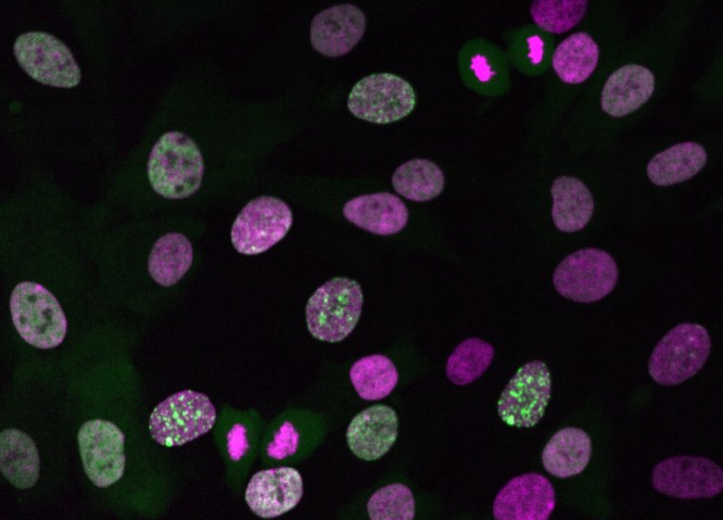
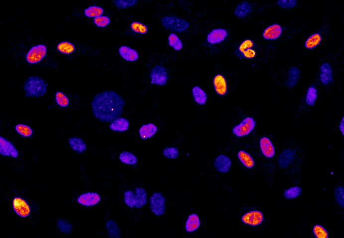
This microscopy image shows living cells at different stages of the cell cycle. Cells with distinct green spots are currently replicating their DNA, which is later packed into chromosomes and segregated to the two daughter cells during cell division.
“Despite the importance of centrosomes for chromosome segregation, centrosomes are lost in the oocytes” explains Dr. Ota. During fertilization, only the sperm brings centrosomes to the newly forming embryo. “When I heard this story as an undergrad, I really wanted to know how the oocytes remove the centrosomes. This is still an enigmatic question” says Dr. Ota.
With their passion for cell cycle research, the researchers of the unit are in good company at OIST, a fact that was important for Dr. Meitinger when choosing to come to Okinawa. “I knew there is already a community of cell cycle researchers here, so it seemed like a great place,” he says and when learning more about the island itself, his excitement for the new location only grew. “I looked OIST up and saw that it was on this small island. Maybe I am also a little bit adventurous,” Dr. Meitinger laughs.
After their first year in Okinawa, both Dr. Meitinger and Dr. Ota look back positively on their experience. “It was a lot of work, but many people helped us set up,” says Dr. Ota. “And I really love the food here!” adds Dr. Meitinger.
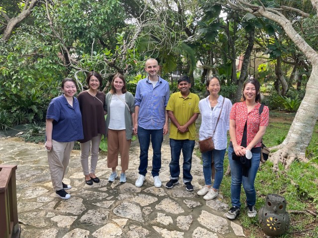
Dr. Midori Ota (second from the right), Dr. Franz Meitinger (fourth from the right), and the Cell Proliferation and Gene Editing Unit Team explore Okinawa during a lunch outing in 2022.
Dr. Midori Ota (second from the right), Dr. Franz Meitinger (fourth from the right), and the Cell Proliferation and Gene Editing Unit Team explore Okinawa during a lunch outing in 2022.
Specialties
Research Unit
Submit a press inquiry


