25 September 2019
Making Okinawa an Imaging Technology Hub

The Imaging Section (IMG) is responsible for maintaining and managing imaging instruments and facility and providing research support at the Okinawa Institute of Science and Technology Graduate University (OIST). Here, sophisticated imaging technologies using photons, electrons, and x-rays are at the service of the scientific community for studies that span from the determination of the three-dimensional atomic structure of proteins to the study of the development of organs in vertebrate species.
In contrast to many other scientific institutions, light and electron microscopy are available in one facility, creating a blend of various techniques from the sample preparation to the imaging of complex specimens, combining the features of fluorescence microscopy with the best spatial resolution delivered by electron microscopy.
The section leader, Dr. Bruno Humbel, has worked at several academic institutions and regularly teaches workshops in different countries. He says he has never seen such an excellent and varied assembly of cutting-edge instruments in a single spot.
The Resources
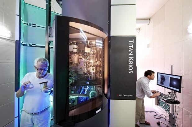
The OIST imaging core facility can count on an impressive array of state-of-the-art instruments that cover most of the applications in light and electron microscopy.
In light microscopy, emphasis is given to systems suitable to image specimens below the fluorescence optical resolution, using known techniques such as PALM, STED and STORM. Fast laser confocal microscopes are also available to image dynamic processes in living specimens. When it comes to imaging large specimens at high-resolution using a non-destructive technology, the IMG makes use of an x-ray microCT scanner.
The structure of biological and material samples can then be imaged at the highest spatial resolution using high-end microscopes — both scanning and transmission electron microscopes. Users have access to three different cryo-electron microscopes. The facility also offers a focused ion beam and serial block-face scanning electron microscope, a high-resolution analytical SEM, and a probe corrected analytical scanning transmission electron microscope with a point resolution of about 80 picometers.
See an image gallery of the equipment.
The section has the instrumentation necessary for sample preparation — especially the cryo-technology for biological electron microscopy.
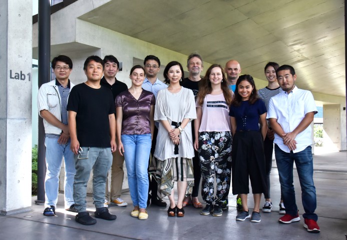
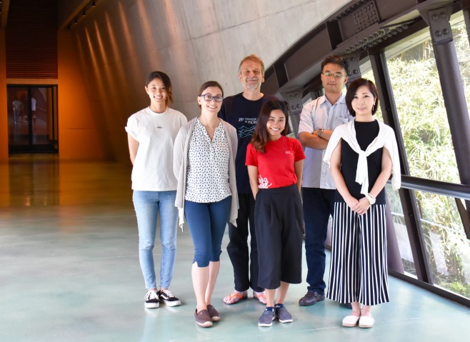
While the in-house technology is valuable, the staff’s skills are priceless. Each member of IMG is a scientist with a longstanding career and expertise in diverse fields. Their experience and knowledge makes the section a gold mine for all fields of science.
IMG offers, in addition to excellent maintenance and user training, their collaboration for scientific projects. This includes helping to design experiments, performing highly sophisticated experiments, project planning, and even assisting with grant applications.
Current Collaborations
The access to the OIST Imaging Section’s resources is open locally, nationally and globally. A local collaboration with three different departments at the University of the Ryukyus has recently been established. The section is also involved with a project at Tokyo University under the auspices of the newly established joint research programme, Jumps Research Collaboration Program (JUMPS), initiated by OIST Provost Dr. Mary Collins.
Dr. Humbel is also engaged in outreach activities to help strengthen OIST’s visibility within the scientific community, and he plays a role in setting up and extending a network of international collaborations with institutions worldwide in the field of electron microscopy.
“Okinawa has a unique position as an island close to many Asian countries,” says Humbel. “My dream is to make OIST, and hence Okinawa, a hub for the Asian scientific community, including Oceania.”
Teaching
To disseminate their expert knowledge, the section is actively involved in teaching and training activities.
The IMG Section is organising annual specialised courses in the field of microscopy such as the “Advanced Light Microscopy Course” under the banner of ABiS. The ABiS programme is a national initiative for Advanced Bioimaging Support funded by the Japanese Society for the Promotion of Sciences Kakenhi Grant. In 2018, ABiS became an official member of the international microscopy network, Global BioImaging (GBI).
Well-known international experts in this field took part at the “Tokuyasu cryo-sectioning and immunolabelling course”, together with Juntendo University, as speakers and instructors. Participation was open to both internal and external applicants.
Another example is the “Cryo Electron Microscopy Course”, organised in collaboration with BINDS (Basis for Supporting Innovative Drug Discovery and Life Science Research) members from several Japanese universities and research institutions. The project bridges OIST to fellow institutes, supporting outside researchers with using the Imaging Section’s valuable cryo-electron microscope to image proteins used in drug discovery and development. The BINDS programme is funded by the Japan Agency for Medical Research and Development (AMED).
The researchers provided an overview of the current state of this Nobel Prize-winning technology, sharing their experiences in demonstrations and hands-on practice. This provided an opportunity for curious young scientists to explore the world of sample preparation, imaging, and data analysis in the context of the BINDS project. This programme has already created four collaborative research projects and will continue until March 2021.
Beyond Academia
The section’s collaborations span beyond the goals of discovery. As OIST Provost Dr. Mary Collins wishes to utilize this gold mine of a resource in more industry collaborations, IMG strives to foster partnerships with the private sector including local companies. Currently, the section is working with a local Okinawan steel company, using its expertise and material science instruments to analyze their various metal alloys.
IMG is also involved in a project with a Japanese company producing synthetic fibres that aims to evaluate the use of sustainable natural materials.
Submit a press inquiry