


Protein Tomography Models

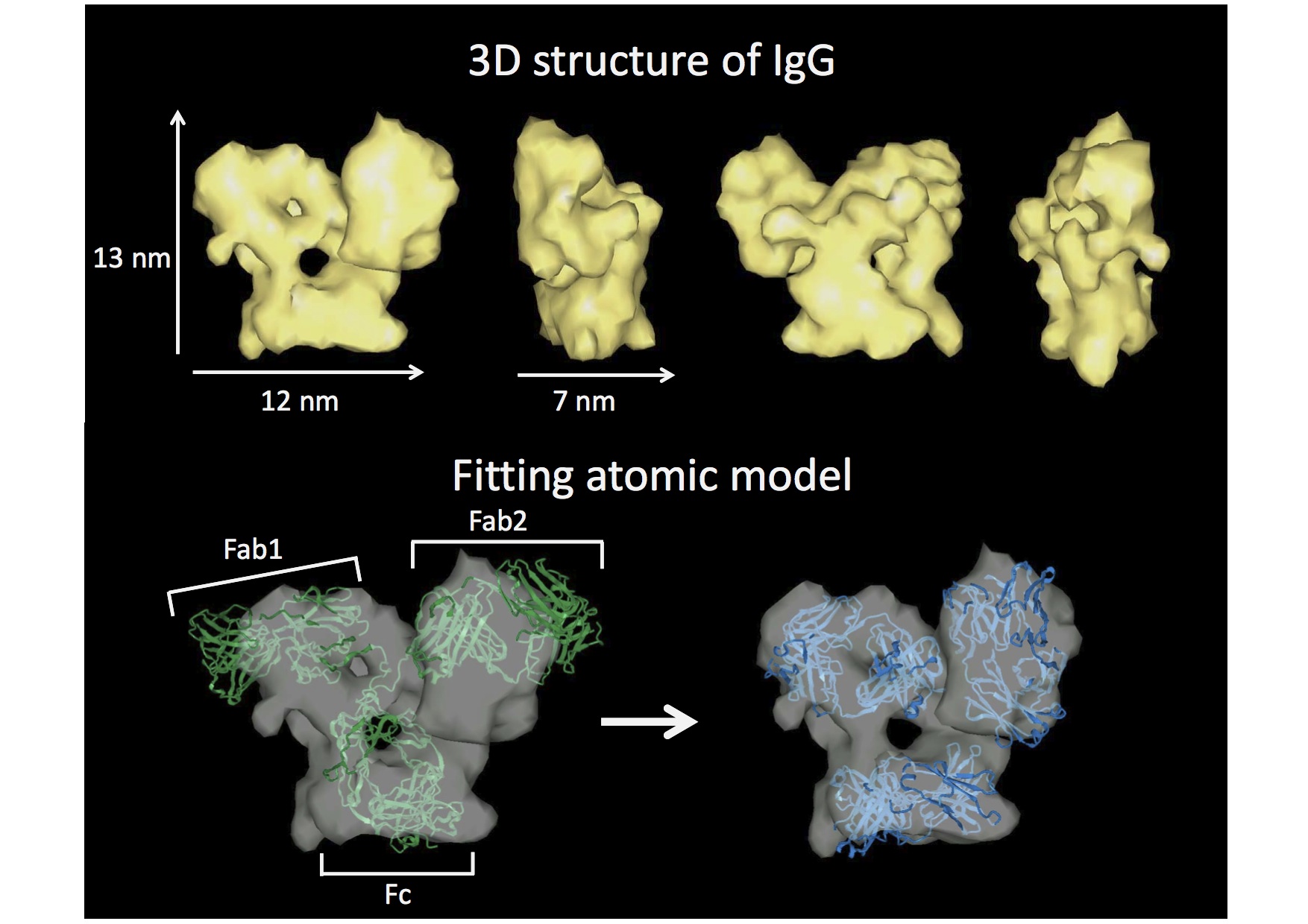
The images show the 3D structure of immunoglobulin using the structural visualizing technique provided by Okinawa Protein Tomography Ltd. (Okinawa PT). In the upper images (yellow), the structure is rotated every 90-degree along the vertical axis. In the bottom two images, the ribbon model obtained through X-ray structural analysis (green and blue) is overlaid with the images. Protein has highly flexible structure and analyzing the structure of individual molecule is essential to understand the function of the protein in vivo. Okinawa PT’s technique has great advantage to analyze the flexibility of protein structure at a single-molecular level.
Copyright OIST (Okinawa Institute of Science and Technology Graduate University, 沖縄科学技術大学院大学). Creative Commons Attribution 4.0 International License (CC BY 4.0).
Tags


