


Unit on Neural Systems and Behavior
Professor Masaki Isoda
Abstract
The goal of this unit is to understand how neurons in both local and global networks implement a variety of higher-order brain functions such as coordinated movement, attention, emotion, and social cognition. Using integrative approaches encompassing single/multi-unit recording, local application of pharmacological compounds, electrical brain stimulation, and development of reversible animal models of cognitive-motor disorders, this unit has clarified (i) the neuronal substrates for self-other distinction in the domain of motor action, (ii) the neuronal architecture and mechanisms underlying social error monitoring, and (iii) pathophysiology and therapeutic mechanisms of basal ganglia mediated motor disorders.
1. Staff
- Dr. Masaki Isoda, Professor
- Dr. Kevin McCairn, Researcher
- Dr. Kyoko Yoshida, Visiting Researcher
- Ms. Akiko Hara , Administrative Assistant
2. Collaborations
- Theme: Neural basis of social cognition
- Type of collaboration: Joint research
- Researchers:
- Dr. Atsushi Iriki, RIKEN Brain Science Institute, Wako, Japan
- Theme: Neural basis of social cognition
- Type of collaboration: Scientific collaboration
- Researchers:
- Dr. Nobuhito Saito, Graduate School of Medicine, The University of Tokyo, Hongo, Japan
- Theme: Cortico-basal ganglia mechanisms of purposive motor behavior
- Type of collaboration: Scientific collaboration
- Researchers:
- Dr. Okihide Hikosaka, National Eye Institute, NIH, Bethesda, U.S.A.
3. Activities and Findings
3.1 Neuronal representation of self-action and others’ action
Successful social interaction depends not only on the ability to identify with others, but also to be able to distinguish between aspects of self and others. Although there is considerable knowledge of a shared neural substrate between self and others in the domain of motor action, the neural mechanism underlying their unique representations remains largely unknown. Exploring such agent-specific mechanisms is important as the proper identification of one’s own actions and others’ actions underlies a number of social behavioral processes, such as learning from others, deciding the optimal level of cooperation and reciprocity, and reading the mental states of others.
To address the issue of how the brain processes self- and others-related behavioral information, we devised an interactive decision task for macaques. This task required two monkeys to monitor each other’s actions for their own action selection. We confirmed that our monkeys were capable of keeping track of the correctness of partner’s actions and utilizing it for determining one’s own actions. Using this behavioral model, we were able to monitor spiking activity of individual neurons in the medial frontal cortex (MFC) – a critical node in the social brain networks in both human and nonhuman primates.
We found that apart from neural activity specifically encoding one’s own actions (self-type), the MFC contained a unique neural code for other’s actions (partner type) and a shared neural code between self-actions and other’s actions (mirror type). The self-type neurons were significantly more prevalent in the ventral subregion of the MFC including the anterior cingulate cortex, whereas the partner-type neurons were more frequently observed in the dorsal subregion including the presupplementary motor area. Interestingly, we seldom found partner-type neurons and mirror-type neurons in a monkey exhibiting a difficulty monitoring the partner’s performance. These findings suggest that the MFC plays an important role in the monitoring of others’ actions for adaptive control of one’s own behavior.
3.2 Frontal cortical mechanisms of social error monitoring
In social settings, one can observe others’ mistakes and learn to avoid and benefit from them. It has been shown that the medial frontal cortex (MFC) processes error information, but the neural architecture and mechanisms underlying social error monitoring remain unexplored.
Using the above-mentioned task in which pairs of monkeys monitor each other’s action for their own action selection, we identified MFC neurons that exclusively responded to other’s errors. These neurons were distributed in two segregated populations: the dorsomedial convexity, where other’s errors were detected, and cingulate sulcus patches, where executive planning for reward benefit from one’s prospective correct action was encoded. These findings indicate that the MFC contains a dedicated circuit for monitoring and utilizing others’ mistakes during social interactions.
3.3 A nonhuman primate model of Tourette syndrome
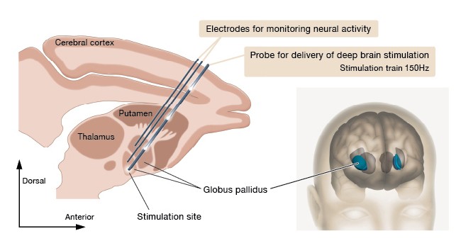
Tourette syndrome is a developmental disorder that is characterized by the occurrence of tics – i.e., sudden, repetitive movements or sounds that people make seemingly without realizing it. Tics range from eye blinking or grimacing to head jerking or foot stamping. Verbal tics can be throat clearing, making clicking sounds, repeated sniffing, yelping, or shouting. Tics are frequently accompanied by other neuropsychiatric disorders such as attention deficit hyperactivity disorder and obsessive compulsive disorder. Previous studies have shown that during the expression of tics, rapid changes in neuronal activity occur in the cerebral cortex and the basal ganglia. Although high-frequency electrical stimulation in the basal ganglia has been tried in clinical trials for Tourette syndrome, a neurophysiological mechanism underlying any therapeutic effect remains a mystery.
To address this issue, we continuously monitored electrical activity of individual neurons in the globus pallidus (GP), a critical element of the basal ganglia, using a nonhuman primate model for Tourette syndrome. We found that during tic expressions, neurons in the external segment of the GP showed a phasic increase in activity, whereas those in the internal segment displayed a phasic decrease in activity. We then applied high-frequency electrical stimulation in the internal segment of the GP with the stimulating parameters mimicking those used in Parkinson’s disease (frequency = 150 Hz, pulse width = 60 µs, intensity = 1 V). To our surprise, deep brain stimulation eliminated abnormal neural activity in more than 70 % of neurons in both segments of the GP. Importantly, these neuronal effects were accompanied by a consistent reduction of movement disturbances. This new research provides a scientific foundation for the clinical application of deep brain stimulation in Tourette syndrome. Future work will build on the current findings toward understanding and treatment of comorbid neuropsychiatric disorders frequently associated with Tourette syndrome.
4. Publications
4.1 Journals
- Isoda M & Hikosaka O. Cortico-basal ganglia mechanisms for overcoming innate, habitual, and motivational behaviors. European Journal of Neuroscience 33: 2058-2069 (2011).
- McCairn KW, Iriki A & Isoda M. High-frequency pallidal stimulation eliminates tic-related neuronal activity in a nonhuman primate model of Tourette syndrome. Neuroreport 23: 206-210 (2012).
4.2 Books and other one-time publications
- 磯田昌岐 & 吉田今日子. 前頭葉における自己と他者の動作表現. Clinical Neuroscience 29: 900-904 (2011).
- McCairn KW, Imamura Y & Isoda M (in press) Animal model of Tics. In Tourette Syndrome (Oxford University Press).
4.3 Oral and Poster Presentations
- Isoda M (2011) Role for the cerebral frontal cortex in processing other’s behavioral information. The workshop on “Neuroscience and economic experiment toward understanding of human society” (Tsukuba, Japan; June 4).
- Isoda M (2011) Representation of self-action and other’s action in medial frontal cortex. Special lecture at Tamagawa University Brain Science Institute (Machida, Japan; August 16).
- Isoda M (2011) The need for fusion research of neuroscience and genome science in nonhuman primates. Workshop on “How to support the future development of neuroscience.” Summer workshop of the Comprehensive Brain Science Network (Kobe, Japan; August 22).
- Isoda M (2011) Neural basis of monitoring self-behavior and other-behavior for strategic decision. PRESTO research meeting. Summer workshop of the Comprehensive Brain Science Network (Kobe, Japan; August 23).
- Isoda M (2011) Monitoring other’s action in the monkey medial frontal cortex. Symposium on “Others in the self: recent advances in social neuroscience.” The 34th Annual Meeting of the Japan Neuroscience Society (Yokohama, Japan; September 15).
- Isoda M (2011) Neuroscience of self and others. Public symposium on “Japanese monkeys bioresource project: achievement in the second term and future perspectives” (Tokyo, Japan; December 9).
- McCairn KW, Imamura Y, Iriki A, Turner RS, Isoda M (2011) The comparative effects of pallidal deep brain stimulation on hypo and hyperkinetic motor disorders in monkeys. The 34th Annual Meeting of the Japan Neuroscience Society (Yokohama, Japan; September 15).
- Isoda M (2012) Neural information processing of one’s own and others’ behavior. NIPS symposium on “Brain’s information processing via global neuronal networks” (Okazaki, Japan; January 7).
- Isoda M (2012) Understanding social cognitive function using Japanese monkeys. NBRP public conference (Tokyo, Japan; January 20).
- Isoda M (2012) Developing a systems neuroscience of self and others using macaques. International symposium on “Frontiers in primate neuroscience researches” (Tokyo, Japan; February 23).
- Isoda M (2012) Basal ganglia (I). RIKEN Brain Science Training Program (Wako, Japan; March 19).
- McCairn KW (2012) Basal ganglia (II). RIKEN Brain Science Training Program (Wako, Japan; March 19).
5. Intellectual Property Rights and Other Specific Achievements
Nothing to report
6. Meetings and Events
Nothing to report
7. Other
Nothing to report.