


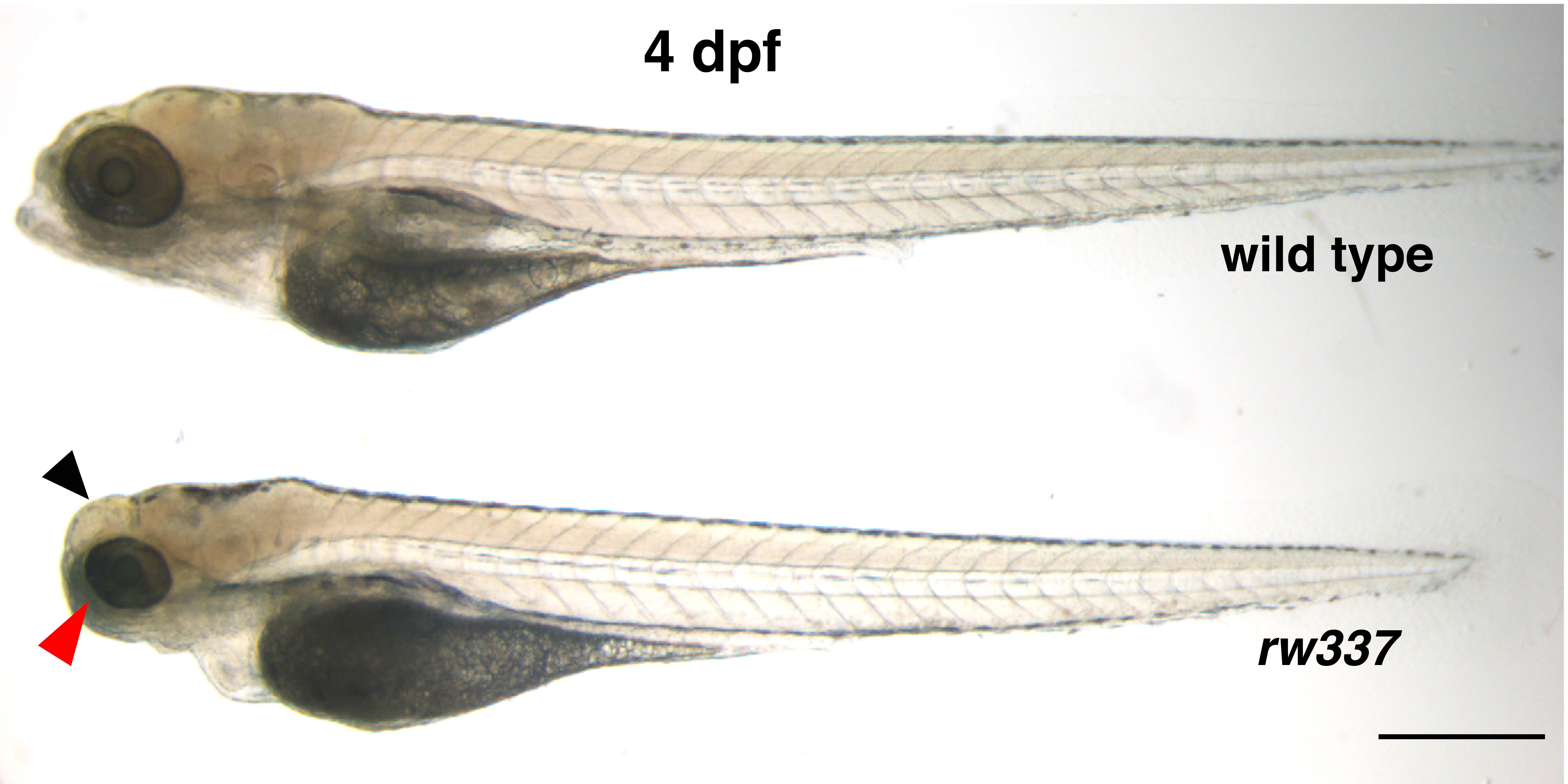
The embryo of a wild type zebrafish above the embryo of banp mutant zebrafish, both four days post fertilization (dpf). The mutant embryo has smaller eyes compared to the wild type (shown with the red arrow). The black arrow shows cloudy tectum (the upper

The embryo of a wild type zebrafish above the embryo of banp mutant zebrafish, both four days post fertilization (dpf). The mutant embryo has smaller eyes compared to the wild type (shown with the red arrow). The black arrow shows cloudy tectum (the uppermost part of the midbrain) in the mutant embryo, which represents cell death. Scale bar = 100 μm. The image appeared in the research paper published in eLife.
The embryo of a wild type zebrafish above the embryo of banp mutant zebrafish, both four days post fertilization (dpf). The mutant embryo has smaller eyes compared to the wild type (shown with the red arrow). The black arrow shows cloudy tectum (the uppermost part of the midbrain) in the mutant embryo, which represents cell death. Scale bar = 100 μm. The image appeared in the research paper published in eLife.
Copyright OIST (Okinawa Institute of Science and Technology Graduate University, 沖縄科学技術大学院大学). Creative Commons Attribution 4.0 International License (CC BY 4.0).


