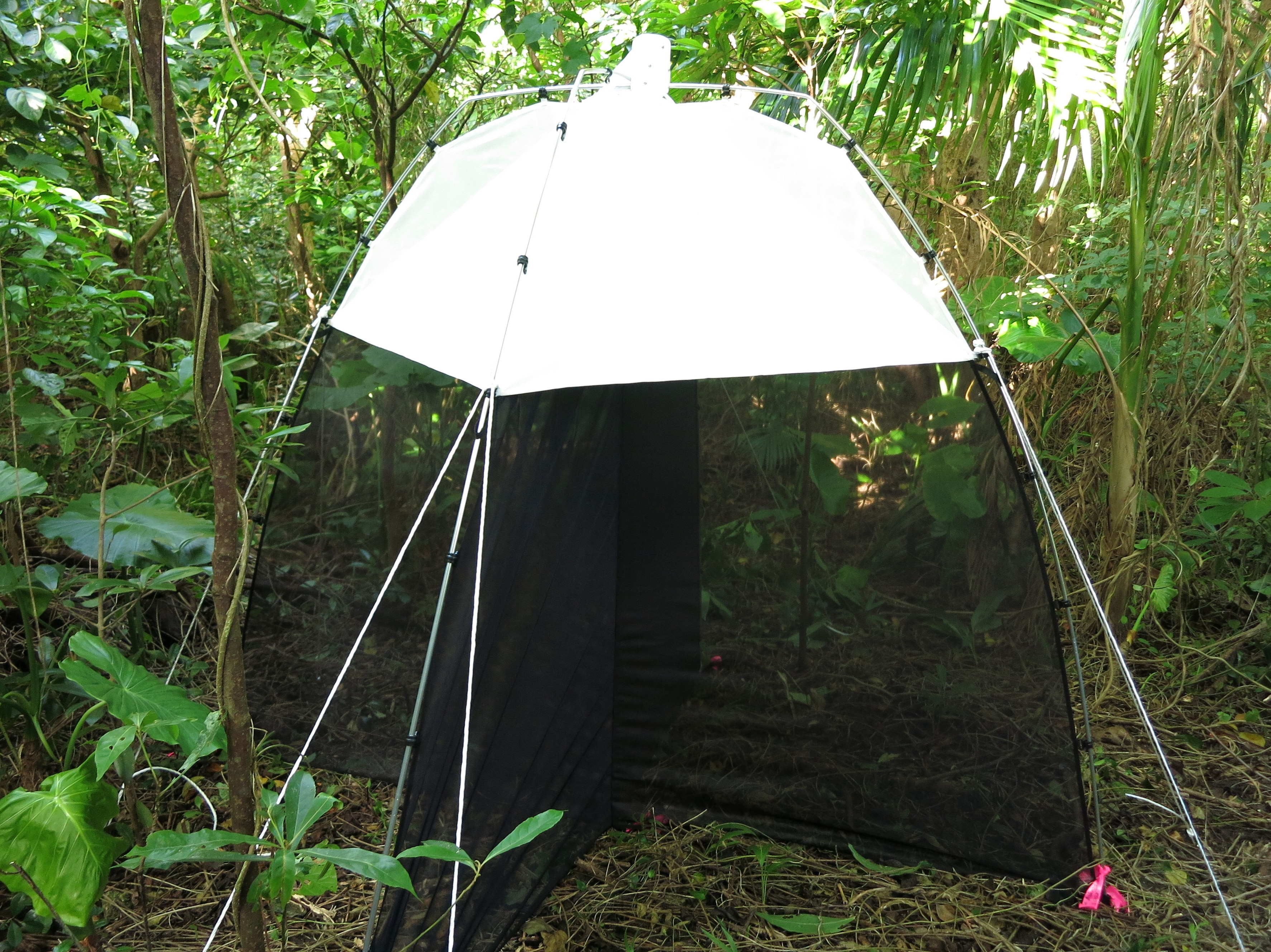
Ants were collected in special traps Ants were collected in special traps at 24 sites across Okinawa over two years. Date: 27 October 2023 OKEON, OIST Download full-resolution image Share on: Related Images Palladium Nanoparticles at Different Temperatures Grammatikopoulos simulated two palladium nanoparticles colliding at different temperatures. The hotter the temperature, the more homogenous the resulting product, and the further the atoms in the particle crystallize. Prof. Ulf Skoglund Prof. Ulf Skoglund Protein Tomography Models The images show the 3D structure of immunoglobulin using the structural visualizing technique provided by Okinawa Protein Tomography Ltd. (Okinawa PT). In the upper images (yellow), the structure is rotated every 90-degree along the vertical axis. In the bottom two images, the ribbon model obtained through X-ray structural analysis (green and blue) is overlaid with the images. Protein has highly flexible structure and analyzing the structure of individual molecule is essential to understand the function of the protein in vivo. Okinawa PT’s technique has great advantage to analyze the flexibility of protein structure at a single-molecular level. Let's make a robot Making a robot using LEGO TC Neuron A thalamocortical, or TC neuron labeled with fluorescent dye, as used in Dr. Augustinaite’s study. The image shows a voltage recording device, at bottom left, entering the yellow cell body, and a stimulation device, at top, reaching the dendrites. Color in this image shows the depth in the slice.
Palladium Nanoparticles at Different Temperatures Grammatikopoulos simulated two palladium nanoparticles colliding at different temperatures. The hotter the temperature, the more homogenous the resulting product, and the further the atoms in the particle crystallize.
Protein Tomography Models The images show the 3D structure of immunoglobulin using the structural visualizing technique provided by Okinawa Protein Tomography Ltd. (Okinawa PT). In the upper images (yellow), the structure is rotated every 90-degree along the vertical axis. In the bottom two images, the ribbon model obtained through X-ray structural analysis (green and blue) is overlaid with the images. Protein has highly flexible structure and analyzing the structure of individual molecule is essential to understand the function of the protein in vivo. Okinawa PT’s technique has great advantage to analyze the flexibility of protein structure at a single-molecular level.
TC Neuron A thalamocortical, or TC neuron labeled with fluorescent dye, as used in Dr. Augustinaite’s study. The image shows a voltage recording device, at bottom left, entering the yellow cell body, and a stimulation device, at top, reaching the dendrites. Color in this image shows the depth in the slice.