


Trans-Membrane Trafficking Unit
Principal Investigator: Fadel Samatey
Research Theme: Functional and Structural Study of the Bacterial Type III Secretion System
Abstract
Bacteria move in their environment by means of flagella. The bacterial flagellum is a long, thin filament extending from the cytoplasm to the cell exterior. All flagellar proteins that reside beyond the cell membrane are exported by the flagellum-specific mechanism that is part of a family of type III secretion systems (T3SS).
In Salmonella typhimurium, the flagellar export apparatus consists of six membrane proteins, FlhA, FlhB, FliO, FliP, FliQ, and FliR, and three cytoplasmic proteins, FliH, FliI, and FliJ. The export apparatus is responsible not only for translocation of flagellar proteins across cell membrane but it also regulates the order in which the proteins are exported. The membrane protein FlhB has been found to play a key role in the regulation process.
This protein is about 40 kDa and consists of two domains: the hydrophobic N-terminal part that is predicted to have four transmembrane helices and the C-terminal cytoplasmic domain. FlhB homologues were found in all type III secretion systems: both flagellar T3SS and T3SS from bacterial needles.
Recently, several different structures of the cytoplasmic domain of FlhB paralogues from bacterial needles have been published. Sequence homology (about 20% of identity and 35% of similarity) between flagellar FlhB and its needle paralogue assumes that general spatial organization may be the same for the two types of the protein. Nevertheless there are also differences. Thus it was shown in our unit that SsaU and SpaS proteins (FlhB paralogues from Salmonella typhimurium needles) couldn't substitute Salmonella flagellar FlhB (C. Barker et al., unpublished observations). To reveal the difference between flagellar FlhB and its needle paralogue we attempted to crystallize and to solve the structure of cytoplasmic domain of Salmonella FlhB and Aquifex aeolicus FlhB. Both protein cytoplasmic domains have been purified and successfully crystallized. Their structures are now being refined.
1. Staff
Researchers: Clive S. Barker, Yasuji Kido, Hideyuki Matsunami, Vladimir Meshcheryakov, Young-Ho Yoon,
Technicians: Paula Bulieris, Julie Carrere, Irina Meshcheyrakova
Research administrator / Secretary: Saeko Hedo
2. Collaborations
Graduate School of Frontier Bioscience, Osaka University, Japan
Theme: Expression and purification of the membrane protein complexes
Type of partnership: Joint research
Name of principal researcher: Prof. Keiichi Namba
Name of researchers: Keiichi Namba, Katsumi Imada
Institute of Laura-Langevin, Grenoble, France
Theme: Molecular dynamic of the bacteria flagellum by neutron scattering
Type of partnership: Collaboration
Name of principal researcher: Dr. Giuseppe Zaccai
CNRS, Institute of Structural Biology and Microbiology (IBSM), Marseille, France
Theme: Structural Study of the bacterial Type II secretion system
Type of partnership: Collaboration
Name of principal researcher: Dr. Jean-Rome Voulhoux
Name of researchers: Jean-Rome Voulhoux, Gerard Michel
Robert Wood Johnson Medical School, New Jersey, USA
Theme: Structural investigation of disordered proteins
Name of principal researcher: Dr. Alla Kostyukova
Research theme: Structural investigation of disordered proteins
Institute of Biologie Physico-Chimique, CNRS, Paris, France
Theme: In vitro, in vivo high-yield production of membrane proteins
Type of partnership: Collaboration
Name of principal researcher : Dr. Bruno Miroux
Name of researchers: Bruno Miroux, Jean-Luc Popot, Francesca Zito
3. Activities and Findings
Flagellum is a huge complex organelle used for motility by many bacteria. Most of the flagellar proteins are localized outside of the cell and are exported across the cytoplasmic membrane by the flagellum-specific secretion apparatus, which shares similarity with the bacterial type III secretion system that is used to secrete effectors into host cell cytoplasm. In Salmonella, flagellum type III secretion system consists of six integral membrane proteins (FlhA, FlhB, FliO, FliP, FliQ, FliR) and three cytoplasmic proteins (FliH, FliI, FliJ). The export apparatus is responsible not only for translocation of flagellar proteins across cell membrane but it also regulates the order in which the proteins are exported. Upon completion of the hook, the secretion system switches substrate specificity from hook-type export to filament-type export. Three proteins, membrane protein FlhB, the hook-length control protein FliK and Flk (also known as RflH or Fluke), play active role in the regulation process.
Salmonella FlhB molecular weight is about 42-kDa and the protein consists of two domains: the hydrophobic N-terminal part, 211 residues, that is predicted to have four transmembrane helices, and the C-terminal cytoplasmic domain (FlhBc), 172 residues. To switch substrate specificity FlhB should get signal from FliK. Cells with defective FliK have very long hook (so called polyhook) without filament attached. Suppression mutations of FlhB that can make the switching even in the absence of FliK have been described. All these mutations lie exclusively in FlhBc assuming an important role of this part of the protein in export regulation.
Aquifex aeolicus is one of the most thermophilic bacteria known representing the deepest phylogenetic branch within the eubacterial evolutionary tree. It is motile and possesses monopolar polytrichous flagella. Salmonella FlhBc and Aquifex FlhBc share 39% sequence identity (Figure 1). However, the two proteins are evolutionarily very distant. Neither fliK nor flk were found in the Aquifex genome, suggesting that the regulation of secretion differs in Salmonella and Aquifex. Comparison of the two proteins may provide us with additional information about the mechanism of FlhB function.
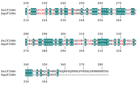
Figure 1. Sequence alignment of FlhBc from S. typhimurium (SalFlhBc) and A. aeolicus (AquFlhBc). Identical residues are shown with a teal background; similar residues are coloured red.
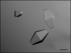
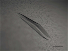
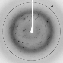
a. Crystallization of FlhBc from Salmonella typhimurium.
Microcrystals of Salmonella FlhBc were obtained in 0.1 M MES pH 6.0, 0.2 M zinc acetate, 40% ethylene glycol. However, the crystals showed no diffraction even using a synchrotron X-ray source. Diffraction-quality crystals were obtained after optimization of each component of the initial crystallization condition. The final crystallization conditions were 0.1 M sodium cacodylate pH 5.0, 0.2 M zinc acetate, 25% propylene glycol. Crystals appeared in 2–3 d after setup of the drop and grew to maximum dimensions of 0.3 x 0.1 x 0.1 mm within a week (Figure 2). The crystals diffracted to 2.45 Å resolution and belonged to space group P42212, with unit-cell parameters a = b = 49.06, c = 142.94 Å, a = b = g = 90.0o.
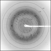
b. Crystallization of FlhBc from Aquifex aeolicus.
Crystals of Aquifex FlhBc were obtained using condition 0.2 M sodium thiocyanate, 20% PEG 3350. The crystals appeared in 3–4 weeks after setup of the drop and grew to maximum dimensions of 0.5 x 0.2 x 0.05 mm within a week (Figure 3). The crystals belonged to space group C2, with unit-cell parameters a = 114.49, b = 33.89, c = 122.13 Å, a = b = 90.0o, g = 107.53 o.
Both structures, SalFlhBc and AquFlhBc, were determined using MAD technique. Refinement of the structures is currently under way.
|
|
Figure 2. Crystals and X-ray diffraction pattern of Salmonella FlhBc. Scale bar, 0.1 mm.
|
|
Figure 3. Crystals and X-ray diffraction pattern of Aquifex FlhBc. Scale bar, 0.1 mm.
4. Publications
4.1 Journals
Barker, C. S., Meshcheryakova, I. V., Kostyukova, A. S., Samatey, F. A., FliO Regulation of FliP in the Formation of the Salmonella enterica Flagellum, 6(9), -ARTN e1001143 DOI 10.1371/journal.pgen.1001143, Plos Genetics
Meshcheryakov, V. A., Yoon, Y. H., Samatey, F. A., Purification, crystallization and preliminary X-ray crystallographic analysis of the C-terminal cytoplasmic domain of FlhB from Aquifex aeolicus, 67, 280-282, Acta Crystallographica Section F Structural Biology and Crystallization Communications, February 2011
4.2 Books and other one-time publications
Nothing to report4.3 Oral and Poster Presentations
Yoshida, S., Matsunami, H., Miyata, T., Kato, T., Namba, K.(Grad. Sch. Front. Biosci., Osaka univ., OIST), Functional domain structure of the bacterial flagellar MS ring, The 10th Annual Meeting of The Protein Science Society of Japan, Hokkaido Japan, June 16-18, 2010
5. Intellectual Property Rights and Other Specific Achievements
Nothing to report6. Meetings and Events
6.1 Seminar
Date: April 2
Venue: New Campus, Lab1 Level C, Meeting room 1
Speaker: Dr. Jean-Luc Popot
Title of the seminar: “Applications of amphipols to membrane protein studies”
Date: April 8
Venue: New Campus, ,Lab1 Level C, Meeting room 1
Speaker: Dr. Eiji Obayashi
Title of the seminar: “Structural insight into essential subunit contacts of the influenza virus RNA polymerase; the basis for new influenza drugs"
Date: August 16
Venue: New Campus, Lab1 Level D, Meeting room 1
Speaker: Dr. Hideyuki Miyatake
Title of the seminar: “A mail-in data collection system and a microfocus beamline at SPring-8”
Date: December 15
Venue: New Campus, Lab1 Level D, Meeting room 1
Speaker: Prof. Simon Silver
Title of the seminar: “Bacterial genes for (almost) all elements of the chemical Periodic Table”
Date: January 14
Venue: New Campus, ,Lab1 Level C, Meeting room 1
Speaker: Dr. Koji Yonekura
Title of the seminar: “Cryo-electron microscopy of biological macromolecular structures”
7. Others
Nothing to report