


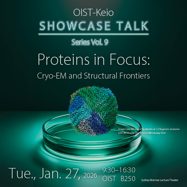
OIST-Keio Showcase Talk Series Vol. 9 - Proteins in Focus: Cryo-EM and Structural Frontiers
Description
Title
OIST-Keio Showcase Talk Series Vol. 9 – Proteins in Focus: Cryo-EM and Structural Frontiers
Symposium Abstract
The ninth edition of the OIST-Keio Showcase Talk Series brings together experts from Keio University, The University of Osaka’s Institute for Protein Research, and OIST to explore cutting-edge advances in protein science and cryo-electron microscopy. Talks will cover drug design, photosynthetic complexes, molecular motors, enzyme evolution, and viral architectures. This symposium highlights how cryo-EM is transforming our understanding of molecular mechanisms and driving innovation in medicine, biotechnology, and environmental science - with the aim of connecting researchers across institutions to enable interdisciplinary innovation.
This symposium will be followed by the 2026 Cryo-Electron Microscopy Course from January 28 to 30, which is aimed at beginners (admission is free). There experienced researchers will provide an overview of the current state-of-the-art and share their experiences in demonstrations and hands-on practice.
Registration
Registration is closed, but walk-ins are welcome. Please come directly to the venue. (Please note that lunch bento will be prioritized for those who ordered in advance.)
Program
*Click here to DOWNLOAD. (As of Jan 26)
The Networking Dinner will be held from 17:30 at OIST's Land neXus.
Keio University Speakers
| Speakers | Affiliation | Talk Title/Abstract | |
| 1 |
Dr. Kunimichi Suzuki Junior Principal Investigator
|
Human Biology Microbiome Kunimichi Suzuki received his BA, MSci and PhD from the University of Tokyo and had a postdoc training in Keio University School of Medicine between 2013 and 2019. He learned the structural biology in MRC-LMB in the UK as an investigator scientist between 2019 and 2025. He also worked as a team leader in the pharma company. In 2025, he started his lab in Keio University Bio2Q. He has worked on the research about the synapse organizing molecules from the structural to behavioral level and now manages the new cryoEM core facility for the versatile biological and medical application. |
Establishment of cryoEM Core Facility and processing pipeline in Bio2Q Cryo-electron microscopy (cryoEM) techniques including single particle analysis (SPA), cryo-electron tomography (cryoET) and microcrystal electron diffraction (MED) has been widely used for multiple biological disciplines and successfully to reveal the molecular structures in a physiological environment. The key element for the successful application of cryoEM techniques to the individual research is highly dependent on the sample quality, performance of cryoEM and the reliable data processing pipeline. We have set up a unique cryoEM core facility in Keio University Bio2Q as an all-in-one lab and here we demonstrate the performance and entire pipeline of the structural analysis. |
| 2 |
Dr. Takanori Yokoo Postdoctoral Scholar
|
Human Biology Microbiome Takanori Yokoo got his Ph.D. at the Department of Chemistry & Biotechnology, Graduate School of Engineering, the University of Tokyo on March 2025. Then he joined Human Biology-Microbiome-Quantum Research Center (Bio2Q) at Keio University in April 2025, and he started to use CryoEM in September 2025. His field of expertise is protein engineering, especially antibody engineering. He has been extensively involved in developing antibody therapeutics targeting synaptic diseases and drug delivery system to brain. |
Structural basis of the complex of the BBB permeable Nanobody and TfR1 Transferrin receptor (TfR1) mediates iron uptake into cells via endocytosis at the blood brain barrier (BBB), making it a promising target molecule for BBB crossing strategies. We aimed to develop Nanobodies (NB) that show BBB permeability. Obtained NB (TO1) bound to mouse TfR1 with high affinity and its BBB permeability has been evaluated in vivo. However, TO1 showed almost no cross-reactivity against human TfR1. To elucidate the tight binding mechanism, the structure of the mouse TfR1-TO1 complex was solved at 2.22 Å resolution using single-particle analysis (SPA). We will discuss the detail molecular mechanism revealed by SPA. |
| 3 |
Dr. Eita Sasaki Senior Assistant Professor
|
Faculty of Pharmacy, Graduate School of Pharmaceutical Sciences Eita Sasaki received his B.S. and M.S. degrees in Pharmaceutical Sciences from the University of Tokyo in 2003 and 2005, respectively. He obtained his Ph.D. in Chemistry from the University of Texas at Austin in 2011. Following postdoctoral research at the ETH Zurich, he joined the Graduate School of Agricultural and Life Sciences, the University of Tokyo, as an Assistant Professor in 2017. Since 2021, he has served as a Senior Assistant Professor at the Faculty of Pharmacy, Keio University. His research interests include the development of fluorescence probes and the engineering of self-assembling proteins. |
Development of a fluorescent protein–fluorescent dye hybrid probe to visualize intracellular molecular crowding The intracellular environment is highly crowded with macromolecules such as proteins and nucleic acids, and this molecular crowding critically influences biomolecular diffusion, interactions, reaction kinetics, and liquid–liquid phase separation. Despite its importance, quantitative and spatial visualization of molecular crowding in living cells remains challenging. In this presentation, we demonstrate that a fluorescent protein–fluorescent dye hybrid molecule, termed Chimera 1–TMR, in which tetramethylrhodamine (TMR) is conjugated to enhanced green fluorescent protein (EGFP) via a HaloTag, has the potential to function as a ratiometric fluorescent probe for imaging intracellular molecular crowding. |
The University of Osaka Speakers
| Speakers | Affiliation | Talk Title/Abstract | |
| 1 |
Dr. Genji Kurisu IPR Director,
|
Institute for Protein Research |
Structural basis of the protein shell facilitating efficient CO₂ fixation in diatom pyrenoids Pyrenoids are subcompartments of algal chloroplasts that concentrate Rubisco enzymes and their CO2 substrate, thereby increasing the efficiency of carbon fixation. Diatoms perform up to 20% of global CO2 fixation, but their pyrenoids remain poorly characterized at a molecular level. In vivo photo-crosslinking to catalogue components of diatom pyrenoids identified pyren2oid shell (PyShell) proteins localized to the pyrenoid periphery of both the pennate diatom, Pheaodactylum tricornutum, and the centric diatom, Thalassiosira pseudonana. In situ cryo-electron tomography (cryo-ET) revealed that the pyrenoids of both diatom species are encased in a lattice-like protein sheath. Disruption of PyShell expression in T. pseudonana resulted in the absence of this protein sheath, altered pyrenoid morphology, and a high-CO2 requiring phenotype, with reduced photosynthetic efficiency and impaired growth under standard atmospheric conditions. Pyrenoids in mutant cells were fragmented and lacked the thylakoid membranes that normally traverse the Rubisco matrix, demonstrating how the PyShell plays a guiding role in establishing pyrenoid architecture. Recombinant PyShell proteins self-assembled into helical tubes and sheets, enabling us to determine a 2.4 Å-resolution PyShell structure. This in vitro structure was fitted into an in situ subtomogram average of the pyrenoid’s protein sheath (Fig.1), yielding a putative atomic model of the PyShell within diatom cells. The structure and function of the diatom PyShell provides a new molecular view of how CO is assimilated in the ocean, a crucial biome that is on the front lines of climate changen1). |
| 2 |
Dr. Takayuki Kato Professor
|
Institute for Protein Research |
Challenges in Visualizing Protein Biogenesis by Cryo-Electron Microscopy In recent years, cryo-electron microscopy (cryo-EM) has undergone a remarkable improvement in achievable resolution owing to the advent of direct electron detectors and advances in image processing methodologies. In contrast to X-ray crystallography, which requires crystallization, and NMR spectroscopy, which is limited by molecular size, cryo-EM enables structural analysis of large and heterogeneous macromolecular assemblies under near-native conditions. This advantage is particularly significant for samples exhibiting structural polymorphism or transient intermediate states, where cryo-EM has emerged as a powerful and often unique experimental approach. In this study, we focused on protein folding processes, especially cotranslational folding that occurs during protein synthesis. The ribosome serves not only as a molecular machine for protein synthesis but also as the initial environment in which nascent polypeptide chains begin to fold. By exploiting the translational arrest sequence SecM, we stalled translation at defined positions and attempted to capture folding intermediates of nascent chains. Although the nascent polypeptide itself is small, the ribosome provides a sufficiently large scaffold to allow high-resolution structural analysis. Using this strategy, we successfully determined ribosome–nascent chain complexes at a resolution of 2.8 Å. The nascent chain within the ribosomal exit tunnel was clearly visualized, whereas regions exposed outside the tunnel became structurally heterogeneous and were not resolved in the averaged reconstruction. We further extended our approach to the bacterial flagellum, a large self-assembling macromolecular machine. Flagellar assembly proceeds via tip growth, in which unfolded component proteins are transported through a narrow central channel and refold at the distal end. To visualize folding and assembly intermediates at the flagellar tip, we fused GFP to the hook protein FlgE. This strategy enabled us to determine the structure of a unique assembly intermediate in which a single FlgE molecule was polymerized, providing structural insight into an early stage of hook construction. In this symposium, we will present the detailed structural features obtained from these two systems and discuss how cryo-EM can be applied to elucidate dynamic processes such as protein folding and self-assembly in large molecular machines. |
OIST Speakers
| Speakers | Affiliation | Talk Title/Abstract | |
| 1 |
Dr. Matthias Wolf Professor
|
Architecture and Contractile Transformation of Myophages Bas63 and ɸTE The BASEL phage collection was developed to provide access to diverse bacteriophages, distinct from model phages. Escherichia phage JohannRWettstein (Bas63), a myophage in the collection, is a member of the Ounavirinae sub-family and the Felixounavirus genus. Using cryogenic electron microscopy, we investigated Bas63’s structure to explore its evolutionary relationships and functional adaptations. Our structures reveal a series of gene products: (1) a capsid decorated with β-tulip proteins at three-fold symmetry axes and a Hoc-like protein at hexamer centers; (2) a conserved connector with an additional 12-fold ring of collar proteins that extend unique whisker proteins that are structurally related to podophage GP4 tail fibers; and (3) a baseplate with long tail fibers resembling a contracted form of T4’s long tail fibers. Sequence conservation analysis of Bas63 structural proteins across ICTV-recognized Felixounavirus’ supports its role as a structural model for Felixounavirus evolution. This study advances the mechanistic understanding of phage architecture and reinforces the structural mosaicism of bacteriophages. AuthorsJames Hodgkinson-Bean1, Rafael Ayala2, Klemens McJarrow-Keller1, Léna Cassin1, Georgia L. Rutter1, Alexander J.M. Crowe1, Matthias Wolf *, 2,3 and Mihnea Bostina*,1,4 1 Department of Microbiology and Immunology, University of Otago, Dunedin, New Zealand 2 Molecular Cryo-Electron Microscopy Unit, Okinawa Institute of Science and Technology Graduate University (OIST), Onna-son, Okinawa, Japan. 3 Institute of Biological Chemistry, Academia Sinica, Nankang, Taipei, Taiwan 4 Theoretical Sciences Visiting Program, Okinawa Institute of Science and Technology Graduate University (OIST), Onna-son, Okinawa, Japan * Corresponding authors |
|
| 2 |
Dr. Oleg Sitsel Assistant Professor
|
Cryoelectron tomography: current state, challenges, future directions Cryoelectron tomography (cryo-ET) is unique amongst structural biology techniques in its capability to visualize protein complexes, cytoskeletal structures, and organelle interactions at up to subnanometer resolution directly in their native cellular environment. This technique has therefore revolutionized our understanding of molecular architecture within cells. However, cryo-ET sample preparation, data acquisition, and downstream processing can be complex. This talk will therefore concentrate on the cryo-ET workflow, highlighting key technical challenges, possible solutions, and future directions of development. |
|
|
3 |
Dr. Prashant Jain Postdoctoral Scholar
|
Minimal Perturbation of Activation Loop Dynamics Rewires Kinase Signaling Enzymes are central to life, with their catalytic activity often shaped by the dynamic conformations of regulatory loops. In hub enzymes such as tyrosine kinases, the activation loop critically controls substrate specificity, catalytic efficiency, and downstream signaling, shaping cellular fate. Yet, the molecular mechanisms by which loop dynamics encode these functions remain incompletely understood. Here, we used SRC kinase as a model to dissect how minimal perturbations of the activation loop reprogram kinase behavior. By generating and characterizing multiple variants, we identified a triple-deletion mutant with altered loop dynamics. Structural and biochemical analyses revealed that this variant explores distinct loop conformations and exhibits a subtle shift in substrate preference toward more acidic motifs. These fine-tuned conformational changes translated into specific cellular signaling outcomes, as demonstrated by phosphoproteomic profiling. Comparative analysis across species further showed that nature exploits similar loop remodeling strategies to modulate kinase function. In this talk, I will highlight how activation loop dynamics can be rationally tuned to reprogram kinase specificity and signaling and discuss the broader implications for rewiring cellular signaling networks. |
Co-hosts
Event News
【Flickr】
【Event Report】 OIST-Keio Showcase Talk Series Vol. 9 Spotlights Cryo EM Breakthroughs and Protein Science Innovation
Contact
Please email academic-partnerships@oist.jp if you have any questions.
※ Please note that this event may be recorded, and the videos may be uploaded by OIST/Keio U. Additionally, photos may be taken during the event for publication online (e.g., the OIST website, social media, etc.). Any non-published information will not be shared. ※Add Event to My Calendar
Subscribe to the OIST Calendar
See OIST events in your calendar app