


Visualizing pH within protein crystals

Proteins constitute the molecular machinery of life. They give structure to cells, pump ions, catalyze reactions, or sense messengers.
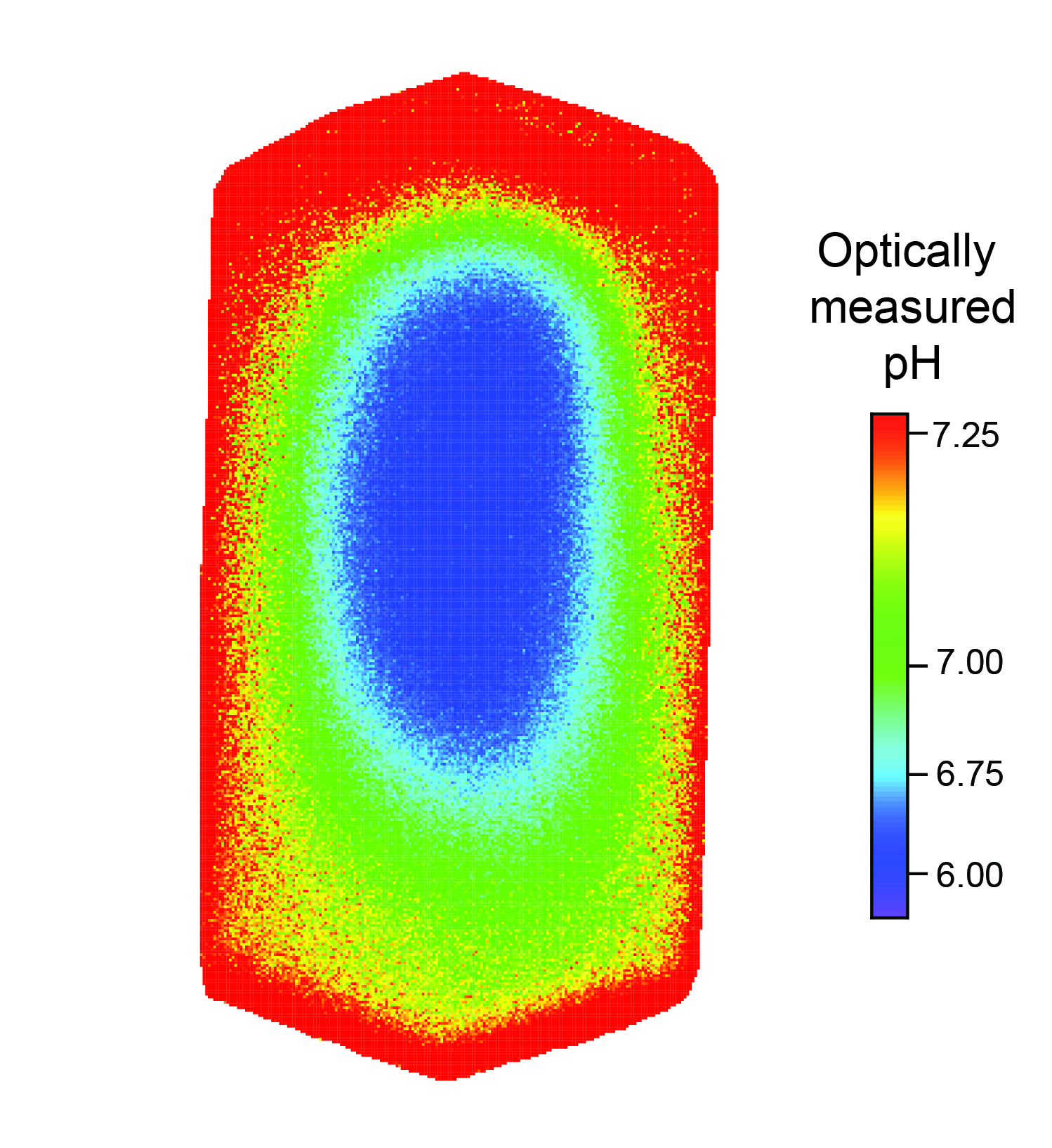
To learn the structure of proteins, researchers coax them into forming a crystal and then use a technique called X-ray diffraction. However, the structure and functionality of proteins is very sensitive to the pH of their surrounding solution, and while the pH in these crystals has been expected to be the same as that of the solution, there has been no method available to confirm this by measuring the pH within protein crystals.
The Optical Neuroimaging Unit developed and published a new optical method that measures the pH within a protein crystal. Interestingly, it turns out that the pH is different from the pH of the bath solution.
Measuring the pH in a crystal is a first step toward changing a crystal’s pH in a defined way. In the future, this might allow researchers to study the structure and function of proteins in crystals at “real-life” pH.
Copyright OIST (Okinawa Institute of Science and Technology Graduate University, 沖縄科学技術大学院大学). Creative Commons Attribution 4.0 International License (CC BY 4.0).
Tags


