Scientists reveal the molecular structure of a complex bacteriophage

The word “virus” is often associated with negative connotations. However, it is important to note that not all viruses are harmful. In fact, there are many viruses that live inside our bodies and play important roles in our health. One example is bacteriophages, viruses that infect bacteria and can be used to keep bacterial infections under control.
These viruses are known to have more complex shapes and have not been studied in full detail at the atomic level before. They can be engineered to better suit applications of human interest, such as providing an alternative to the use of antibiotics.
Scientists at the Okinawa Institute of Science and Technology (OIST) together with their international collaborators at MSU Moscow and Shenzhen, and Academia Sinica in Taiwan, have studied the molecular architecture of the Tequintavirus (DT57 bacteriophage), also known as T5-like bacteriophages, to understand how these viruses are organized at a molecular level. T5 viruses are nonenveloped viruses with a head that has an icosahedral shape and contains the viral DNA, and a non-contractile flexible tail which acts as the channel for DNA injection into the bacterial host cell.
The scientists obtained atomic models for all structural components of the virus. This is the first time that a tailed virus with a flexible tail has been revealed in its entirety at this level of detail. The results of their study have been published in the journal Nature Communications and set the basis for future studies on the mechanism of infection of these viruses.
“To engineer and modify these viruses efficiently for specific purposes, we must know their organization at an atomic level and the mechanisms through which they infect their target bacteria. For these reasons, we decided to use cryo-electron microscopy to visualize the DT57C bacteriophage at high-resolution in its entirety,” Prof. Matthias Wolf, head of the Molecular Cryo-Electron Microscopy Unit, explained.
Researchers working on phage therapies which use bacteriophages to treat bacterial infections in agricultural crops, fish aquaculture and other fields, can benefit from the results of this study. “The structural knowledge we have obtained can enable the engineering of bacteriophages with improved ability to kill these bacterial pathogens,” Prof. Wolf added.
Does this mean that bacteriophages are ‘good’ viruses? Dr. Rafael Ayala, lead author of the research paper, explained that these viruses are ‘good’ when their actions benefit us and ‘bad’ when they cause us harm, as is the case with bacteria.
An example of how bacteriophages can benefit us is their use in gene therapy. “One of the ways to distribute genes to cells is to put them into a human virus that has been modified in two ways, first to not cause disease, and second to also carry the genes that you want to introduce to cure a specific disease. In this way the virus is used as a vehicle to introduce a cure,” Dr. Ayala said.
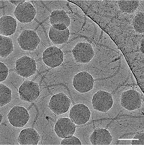
One of the main challenges of the research was to reconstruct the bacteriophage as a whole from electron micrographs in significant detail, and not just some of its components. The DT57C bacteriophage comprises a head, a neck, a tail and a baseplate at the end of the tail. Many of these components are flexible and can move freely, which makes it difficult to visualize their molecular architecture in detail, similar to how difficult it is to take a good photo of an object that is moving fast.
To deal with this, the researchers developed new methods that they plan to apply to other viruses with complex shapes. “We had to think of new ways to tackle the problems we encountered, and we believe that the methods developed in this study will be of interest to many researchers studying viruses,” Dr. Ayala explained. “Phage therapy is an active area of research, and it is very likely that we are going to see these treatments in our lifetime.”
Using viruses to modify bacteria is a huge area of interest because bacteria are at the core of many natural and engineered processes, including nutrient recycling, symbiosis, bioremediation (bacteria are used to clean up environmental pollutants), and food production. This research will be useful in designing viruses to combat bacterial diseases that affect humans, plants, and other organisms.
Article Information
Specialties
Research Unit
For press enquiries:
Press Inquiry Form