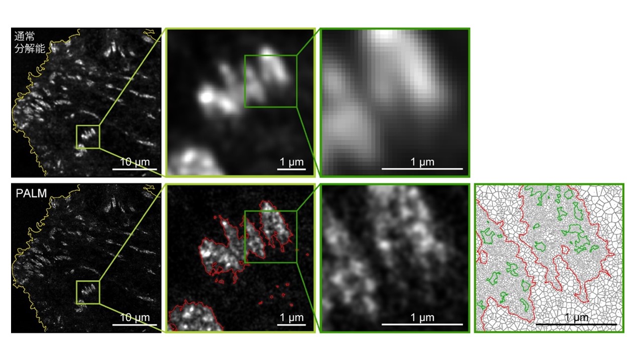
図2 図2 細胞膜を、ほぼ細胞全体にわたって撮影した画像(左端)。接着斑の部分を、順次拡大している(中、右)。上段は、通常分解能の画像。下段は点描法による超解像PALM画像。見ている分子は、接着斑を構成するパキシリン。下段の右端は、すぐ左の画像を定量解析したもの。緑が島の輪郭。赤が接着斑全体の輪郭。 Date: 26 May 2023 Copyright OIST (Okinawa Institute of Science and Technology Graduate University, 沖縄科学技術大学院大学). Creative Commons Attribution 4.0 International License (CC BY 4.0). Download full-resolution image Share on: Related Images Students from Gushikawa High School OIST aims to have every single high school student in Okinawa, like these students from Uruma City, visit the university before they graduate. ROV and xenophyophores A giant foraminifera called a xenophyophore appear as tiny dirt clods next to the robotic arm that collects them. Photo courtesy of JAMSTEC Xenophyophore An 8 cm large xenophyophore specimen from Ogasawara Trench showing clear growth rings. Photo courtesy of Beatrice Lecroq Warm or Cold? Brain slices of older tissues prepared with ice-cold (blue) and warm (red) preparation methods, as demonstrated by Shiwei Huang and Marylka Yoe Uusisaari in a paper in Frontiers in Cellular Neuroscience. The asterisks (*) indicate visible neurons. While no healthy neurons are visible in the left image, seven are visible in the right. Abbreviations: PN, Purkinje neuron layer; ML, molecular layer; GrC, granular cell layer. Shiwei Huang and Marylka Yoe Uusisaari Shiwei Huang (right) is a member of the Computational Neuroscience Unit and Marylka Yoe Uusisaari (left) is a part of the the Optical Neuroimaging Unit.
Students from Gushikawa High School OIST aims to have every single high school student in Okinawa, like these students from Uruma City, visit the university before they graduate.
ROV and xenophyophores A giant foraminifera called a xenophyophore appear as tiny dirt clods next to the robotic arm that collects them. Photo courtesy of JAMSTEC
Xenophyophore An 8 cm large xenophyophore specimen from Ogasawara Trench showing clear growth rings. Photo courtesy of Beatrice Lecroq
Warm or Cold? Brain slices of older tissues prepared with ice-cold (blue) and warm (red) preparation methods, as demonstrated by Shiwei Huang and Marylka Yoe Uusisaari in a paper in Frontiers in Cellular Neuroscience. The asterisks (*) indicate visible neurons. While no healthy neurons are visible in the left image, seven are visible in the right. Abbreviations: PN, Purkinje neuron layer; ML, molecular layer; GrC, granular cell layer.
Shiwei Huang and Marylka Yoe Uusisaari Shiwei Huang (right) is a member of the Computational Neuroscience Unit and Marylka Yoe Uusisaari (left) is a part of the the Optical Neuroimaging Unit.