


onu FY2018 Annual Report 2

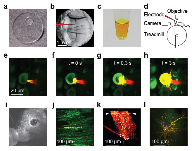
Double-labelling individual neurons for combined voltage and calcium two-photon imaging in awake mice. (a) 5-mm glass cover slip with silicone access port (Roome & Kuhn 2014). (b) A chronic cranial window with access port on the vermis of the cerebellum allows access to the brain with a pipette (schematically indicated). (c) ANNINE-6plus dissolved in pure ethanol at 3 mM concentration. (d) Sketch of the setup with a mouse mounted on a treadmill under a two-photon microscope. An electrode is used to fill single neurons by electroporation and to electrically record from their soma. A behavioral camera allows detailed observation of the pupil, the vibrissa, and the face of the mouse. (e-h) A patch pipette filled with ANNINE-6plus/ethanol solution is used to label single GCaMP6f expressing neurons by electroporation in the anesthetized mouse. (i) During the imaging experiment the mouse is fully awake, sitting on a treadmill and monitored with behavioral a camera. 24 hours after labelling a Purkinje neuron with ANNINE-6plus, the dye has spread out evenly, as can be seen in (j) the cross section of the Purkinje neuron dendrite as an overlay of the green channel (GCaMP6f) and the red channel (ANNINE-6plus) and in (k) the reconstruction of the Purkinje neuron in the red channel (ANNINE-6plus). The dotted line indicates the line scan position used in Fig. 4 and 5. It is also possible to fill other neurons with ANNINE-6plus by electroporation, as, for example, (l) cortical layer 2/3 pyramidal neurons shown as overlaid z-projection of the green channel (GCaMP6f) and the red channel (ANNINE-6plus).
Copyright OIST (Okinawa Institute of Science and Technology Graduate University, 沖縄科学技術大学院大学). Creative Commons Attribution 4.0 International License (CC BY 4.0).


