


onu FY2013 Annual Report 11

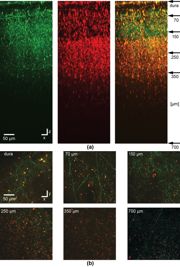
Figure 9: In vivo imaging of NOPS-labeled magnetic nanoparticles or clusters of particles in the barrel cortex of an anesthetized mouse. To label a subset of neurons in barrel cortex, at first, a viral vector is injected delivering the DNA of the fluorescent protein eGFP. 4 days later, magnetic FePt nanoparticles were injected and imaged through a chronic cranial window with multi-photon microscopy in the mouse barrel cortex. A stack of images was recorded with two channels starting from the dura and then reconstructed to show the side view (a). Clusters of NOPS-labeled magnetic FePt core-shell nanoparticles and maybe also single particles are visible in the green (left) and red (middle) channel while the axons and dendrites of eGFP expressing neurons are only visible in the green, as can be seen in the overlay (right). Particles or clusters of particles can be detected in xy images between axons (majority of processes) and dendrites down to a depth of 700 µm (b). Depth of imaging is indicated in the upper left corner corresponding to the arrows in (a). For the image at 700 µm in (b) the contrast and averaging was increased in comparison to the images at lower depth. The dura is visible due to backscattering of the second harmonic signal of collagen. The excitation wavelength was 1000 nm.
Copyright OIST (Okinawa Institute of Science and Technology Graduate University, 沖縄科学技術大学院大学). Creative Commons Attribution 4.0 International License (CC BY 4.0).


