


nmcpu_oist_nmcpu_fy2020_Figure2

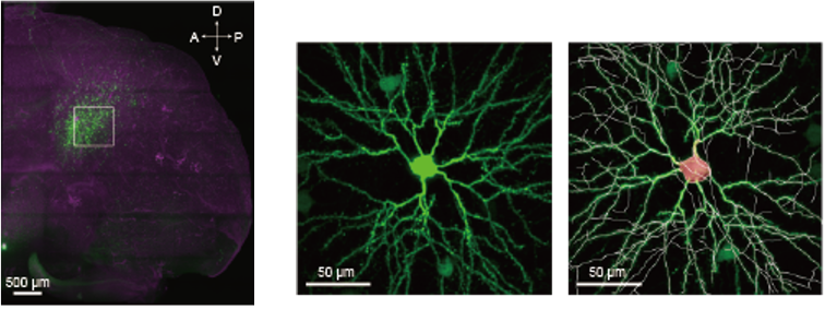
Figure 2: Volumetric imaging of primary auditory cortical neurons by expressing GFP with viral vectors (Left). GFP expressed L3 neurons in a 200 µm subvolume (Middle) and its’ morphological soma (red) and dendrite (white) modeling (Right).
Date:
13 March 2024
Copyright OIST (Okinawa Institute of Science and Technology Graduate University, 沖縄科学技術大学院大学). Creative Commons Attribution 4.0 International License (CC BY 4.0).
Share on:


