


mbnu FY2019 Annual Report 3.1.3

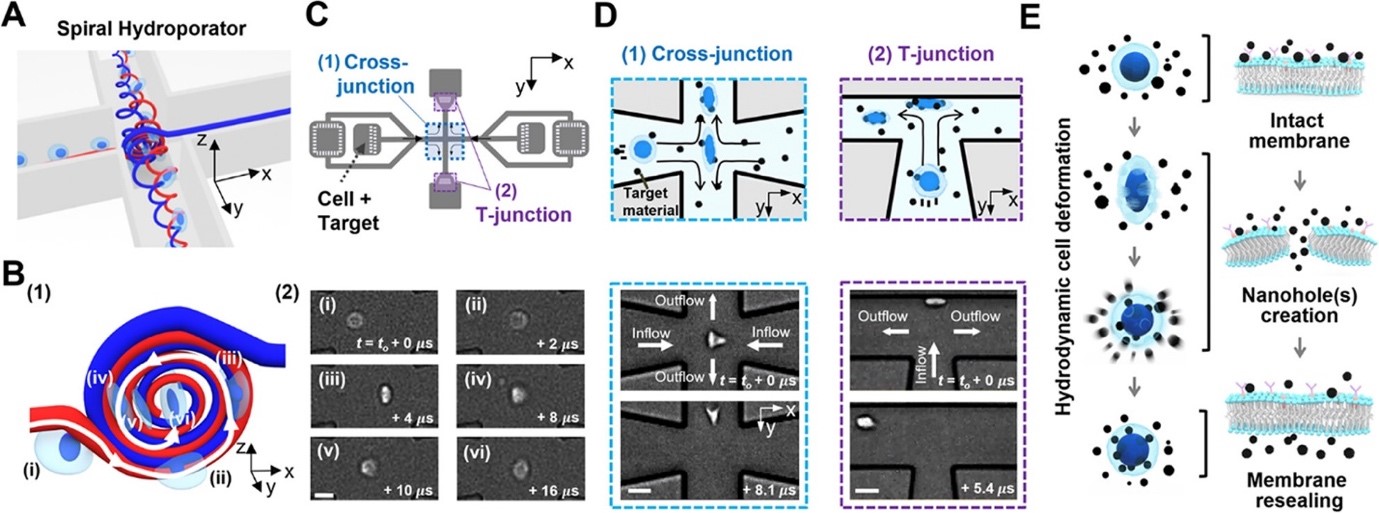
Figure 3: Spiral hydroporation for intracellular nanomaterial delivery. (A) Schematic depicting spiral flow motion at a cross-junction channel. (B) (1) Illustration of spiral vortex-induced cell deformation and (2) high-speed microscope images showing rotational cell motions (scale bar: 10 μm). (C) CAD layout of the hydroporation microfluidic device, consisting of (1) a cross-junction and (2) two dividing T-junction channels. (D) Hydrodynamic cell deformation via (1) the spiral vortex at the cross-junction and (2) cell-wall collision at the T-junction(s) (scale bars: 20 and 30 μm for (1) and (2), respectively). (E) Schematic illustrating nanomaterial delivery into cell cytosols.
Copyright OIST (Okinawa Institute of Science and Technology Graduate University, 沖縄科学技術大学院大学). Creative Commons Attribution 4.0 International License (CC BY 4.0).


