


figure 1

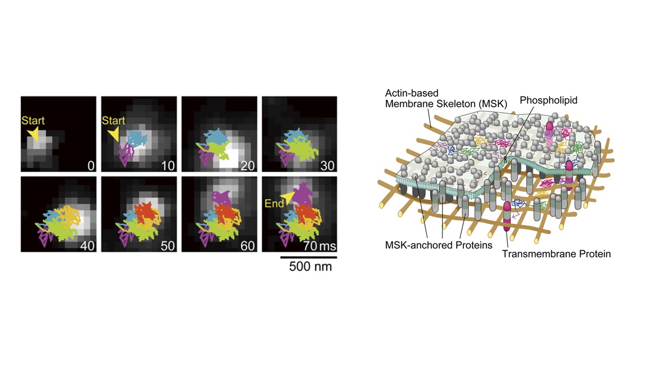
(Left) A representative image sequence of a single Cy3-DOPE molecule diffusing in the apical PM, recorded every 0.1 ms (shown every 100 image frames = every 10 ms). The colors in the trajectory indicate the diffusion in different plausible. (Right) Schematic drawing showing that membrane molecules undergo hop diffusion in the PM, which is compartmentalized by the actin-based membrane-skeleton (MSK) meshes (fences; brown mesh) and rows of transmembrane-protein pickets anchored to and aligned along the actin fence (gray molecules).
Date:
07 June 2023
Copyright OIST (Okinawa Institute of Science and Technology Graduate University, 沖縄科学技術大学院大学). Creative Commons Attribution 4.0 International License (CC BY 4.0).
Share on:


