


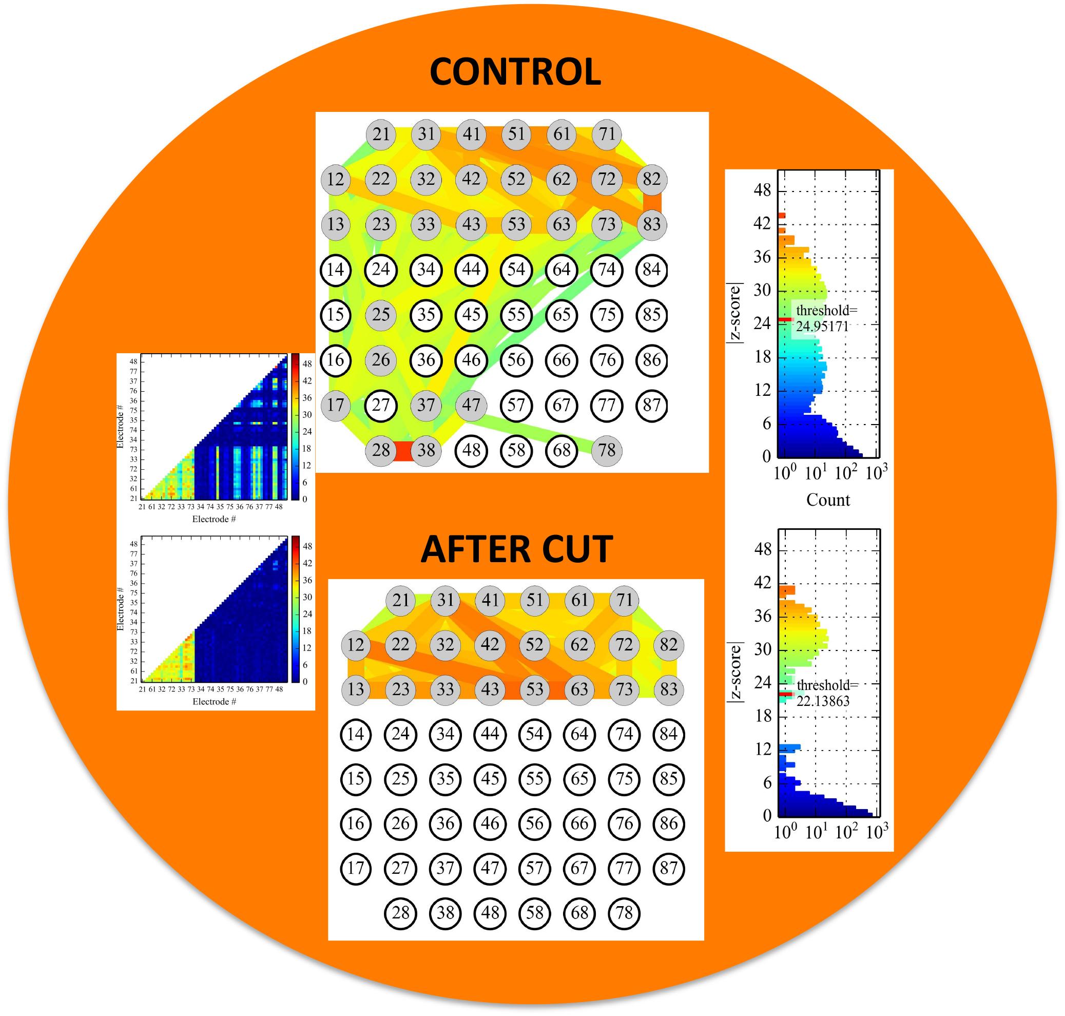
Connectivity map of a Multi-electrode Array (MEA) showing electrophysiological activity between the electrodes.

The top half of the MEA housed the compartment containing cells from the mouse cortex. The bottom half housed the compartment containing cells from the mouse striatum. When the axons reaching across the divide were cut with a knife, the interconnectivity between the cortical neurons inside their own compartment was relatively unaffected, whereas the striatal neurons showed no electrophysiological activity. This demonstrates that there is no back talk from the striatal side to the cortical side in this system and a working corticostriatal network similar to the one inside living brains has been recreated.
The colors are a sign convention used to grade the strength of connectivity between two electrodes regardless of how far apart they are. Blue indicates the weakest connections while green, yellow, orange and red are used, in order, to show stronger and stronger gradations of connectivity.
Copyright OIST (Okinawa Institute of Science and Technology Graduate University, 沖縄科学技術大学院大学). Creative Commons Attribution 4.0 International License (CC BY 4.0).
Tags


